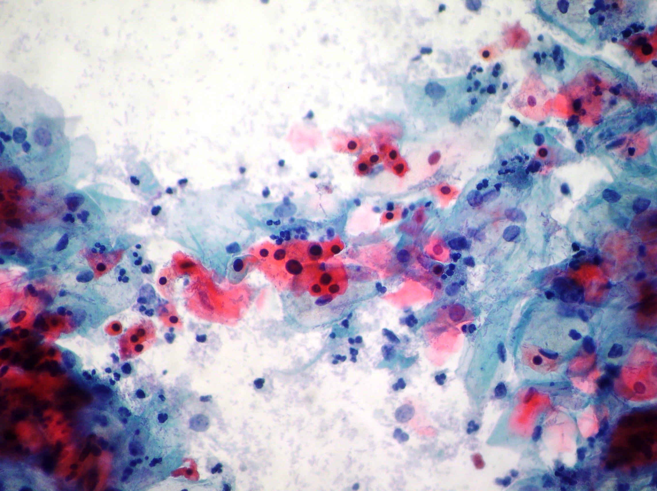
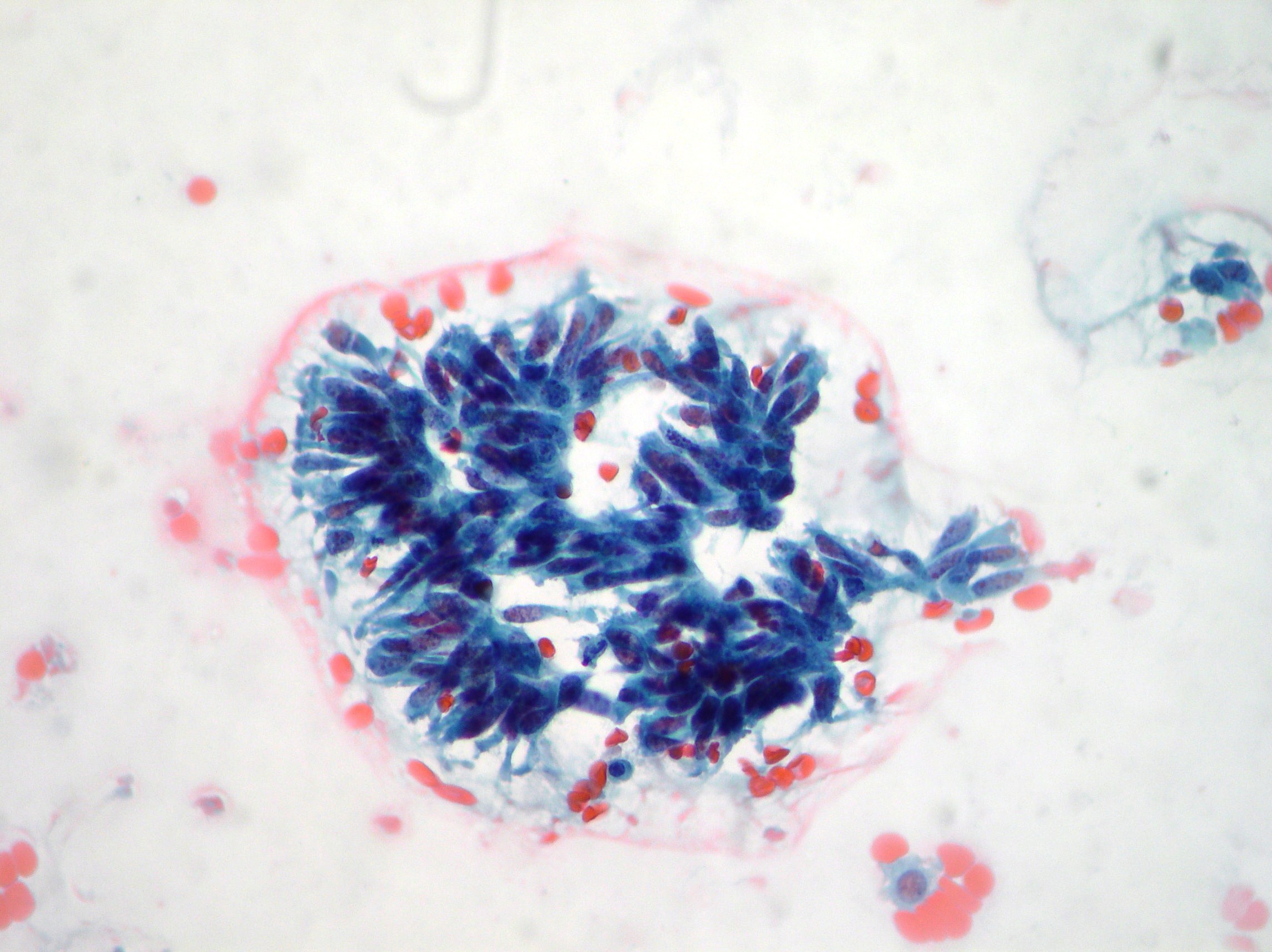
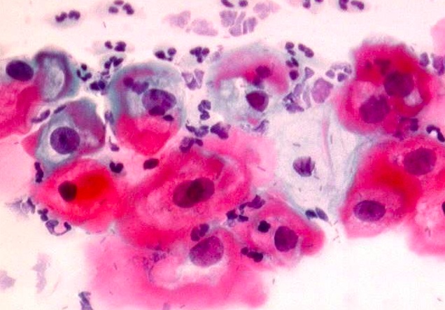
Keratinizing squamous cell carcinoma
Elongated and bizzarre shaped cells, degenerated round and oval nuclei with dense eosinophilic cytoplasm. Inflammation, debris and blood cells in the background. (Papanicolaou, x200, x100)
HSIL
Double staining for p16(INK4a) antigen in a case of HSIL (Thin Prep, x200)
LSIL
HPV infection show typical koilocytotic atypia exhibiting perinuclear halos, condensation of the chromatin and binucleations. (Papanicolaou, x200)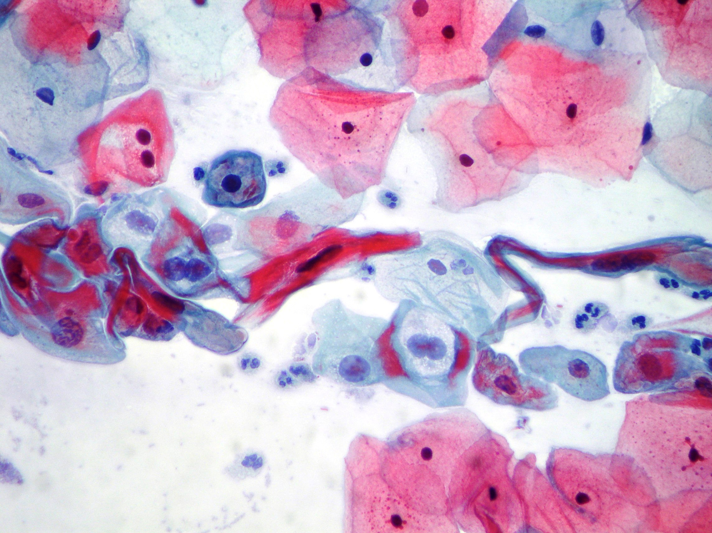
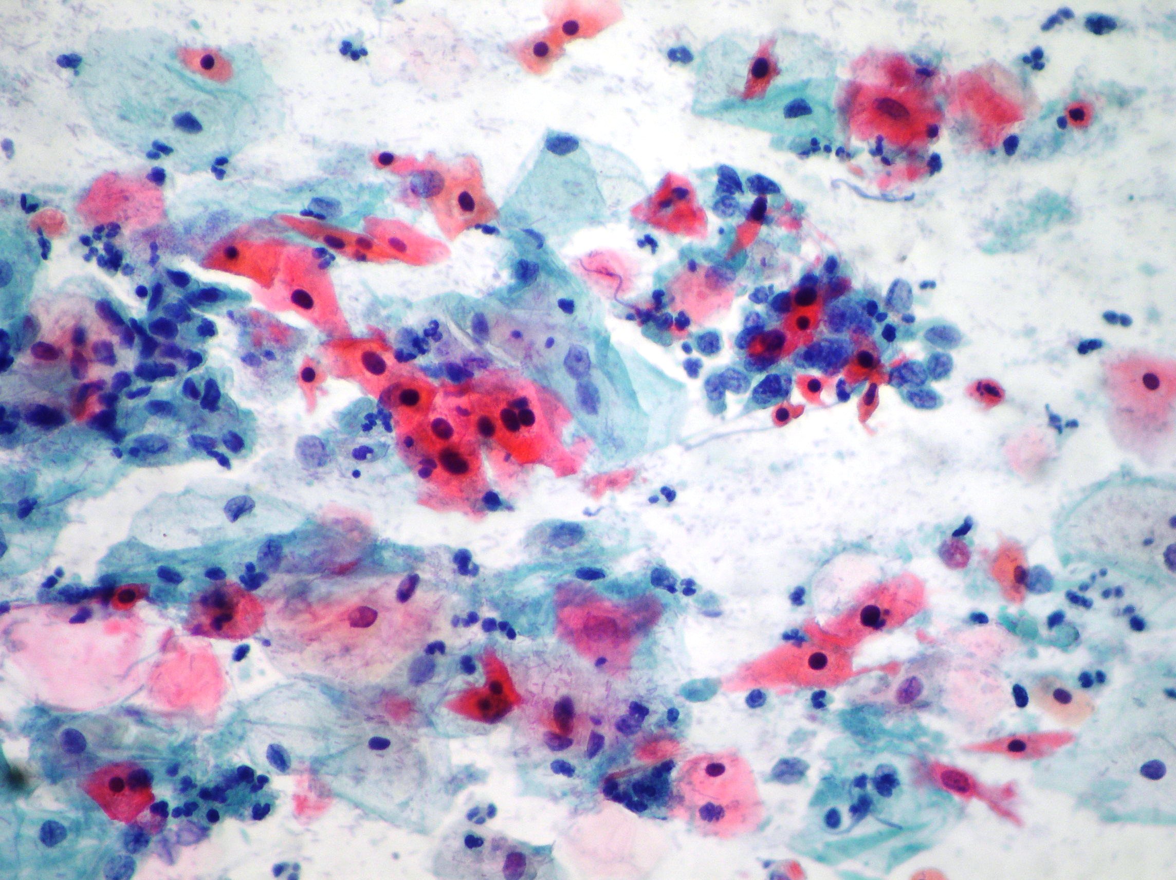
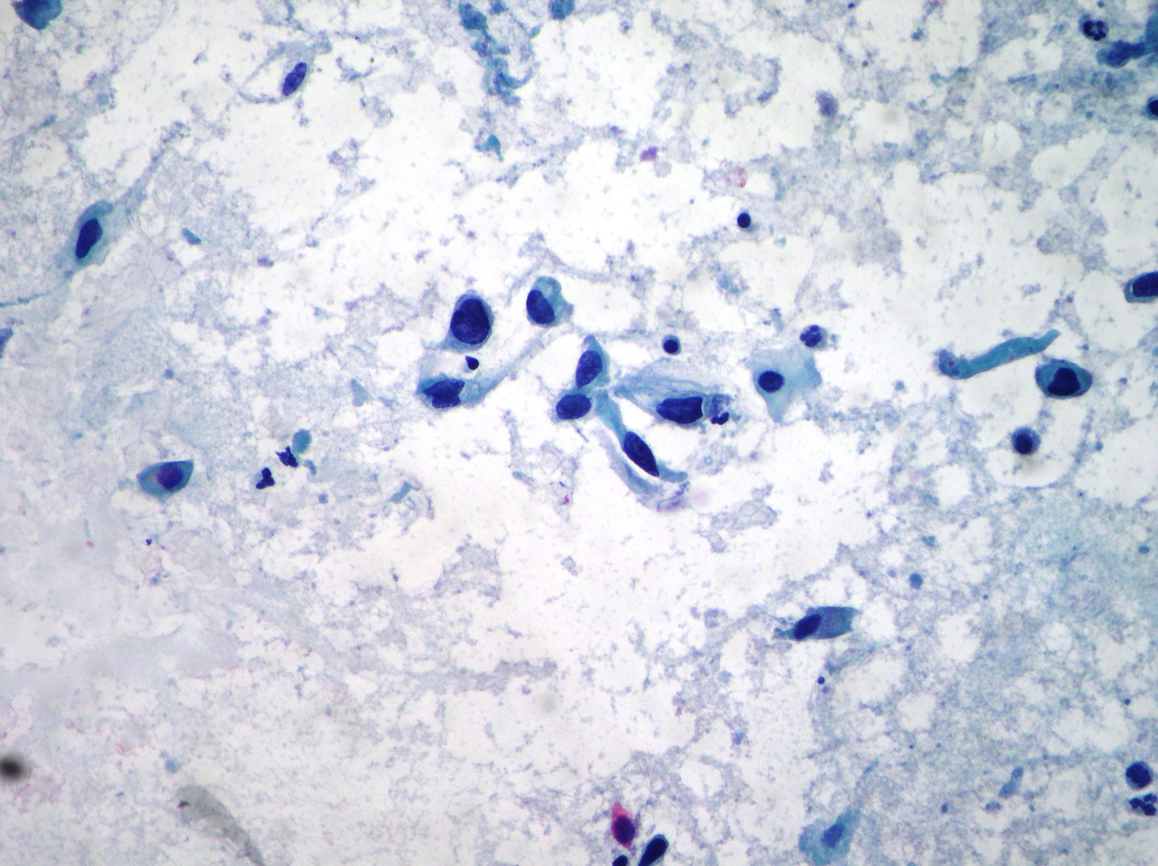
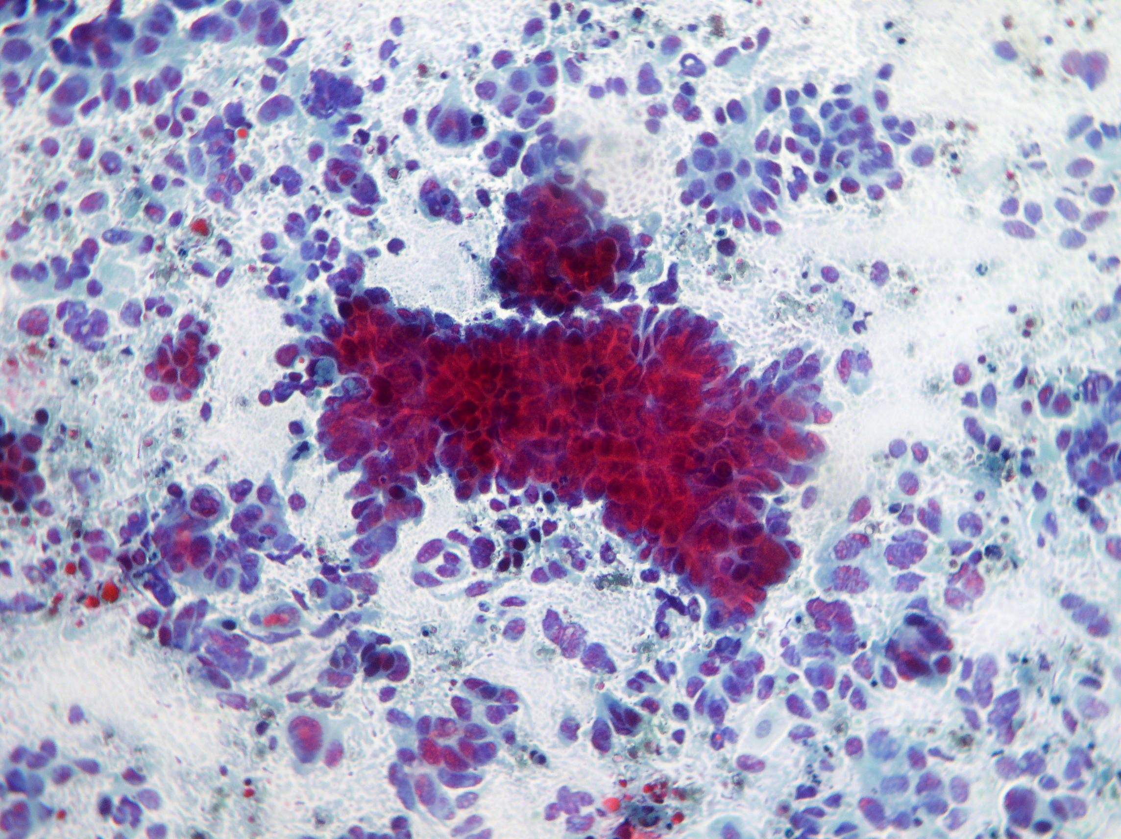
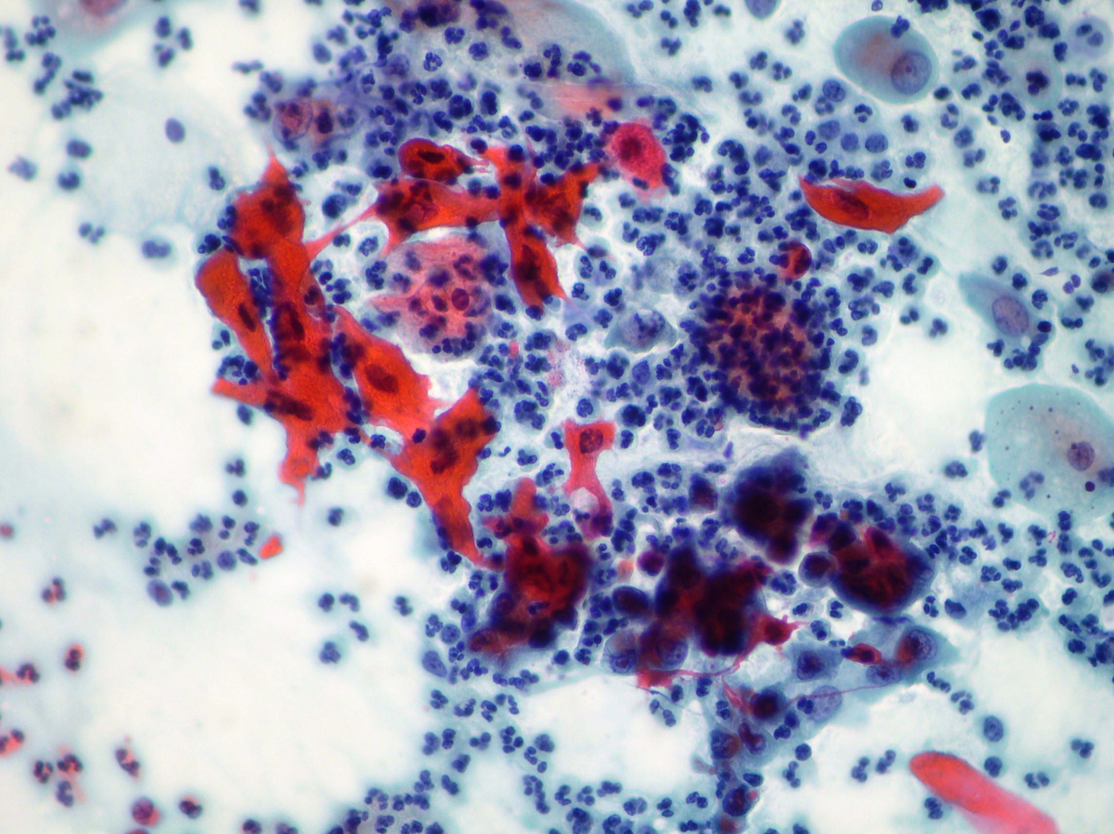
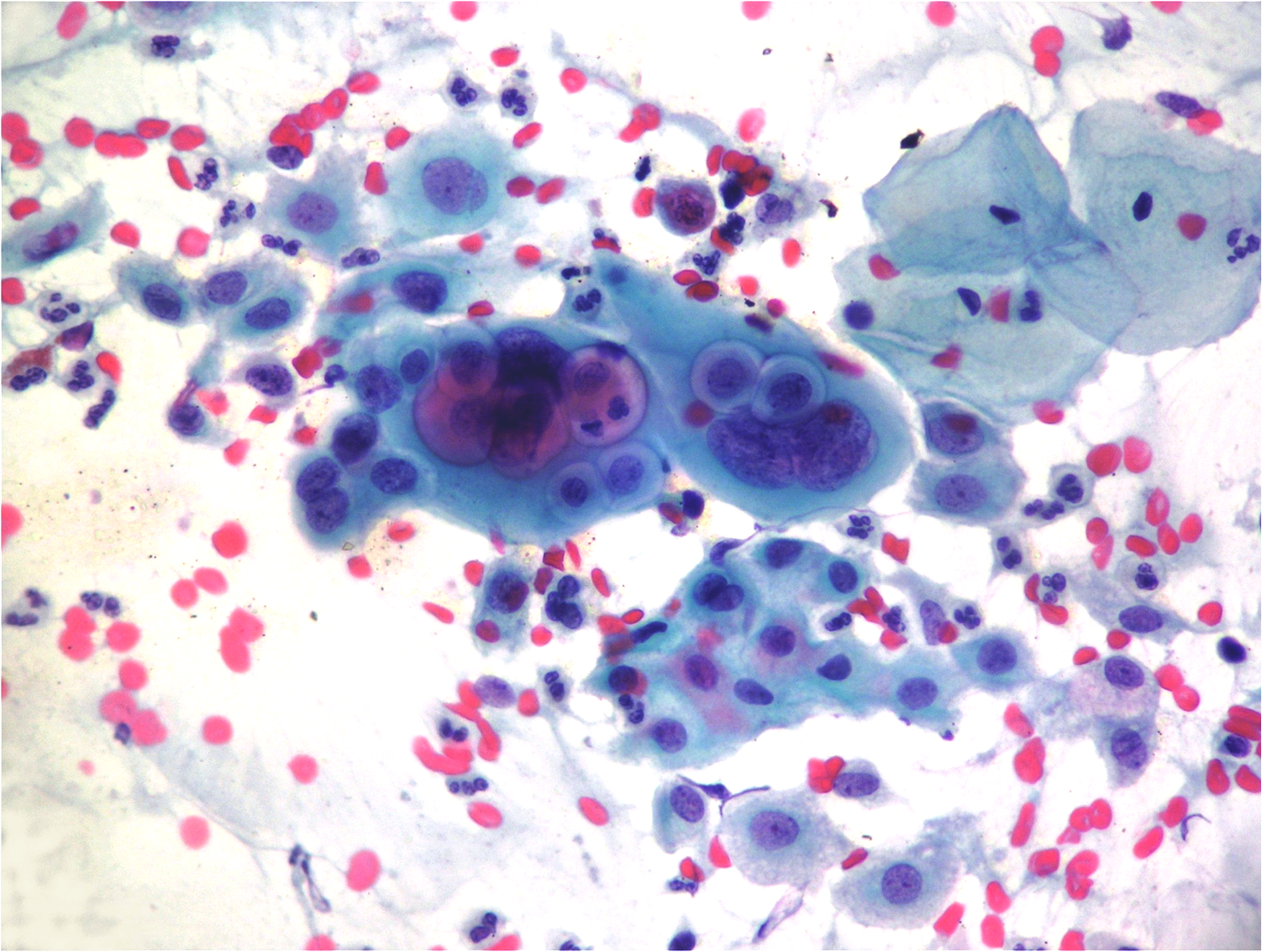
Uterine cervix adenocarcinoma
Cancer cell population showing irregular aggregates with elevated disorganization and individual cell scattering (Papanicolaou x 200)
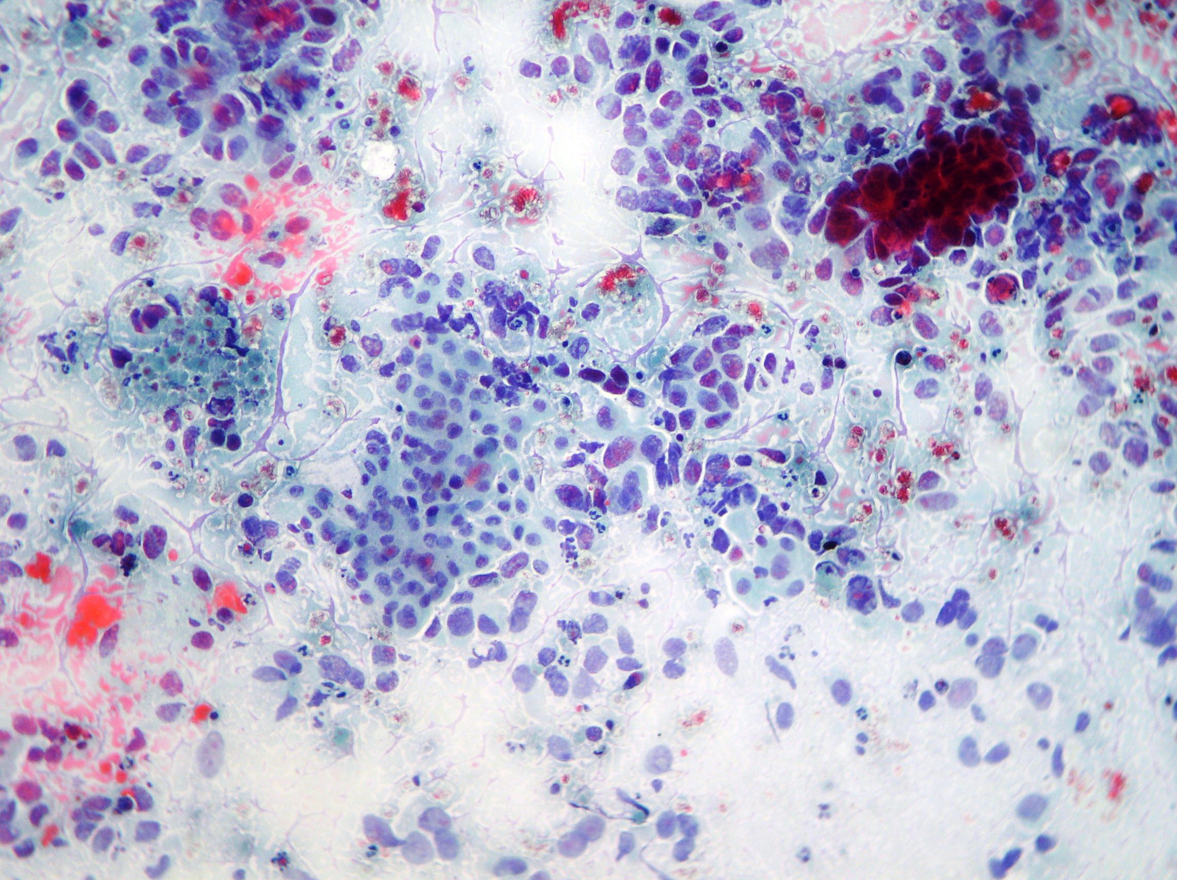
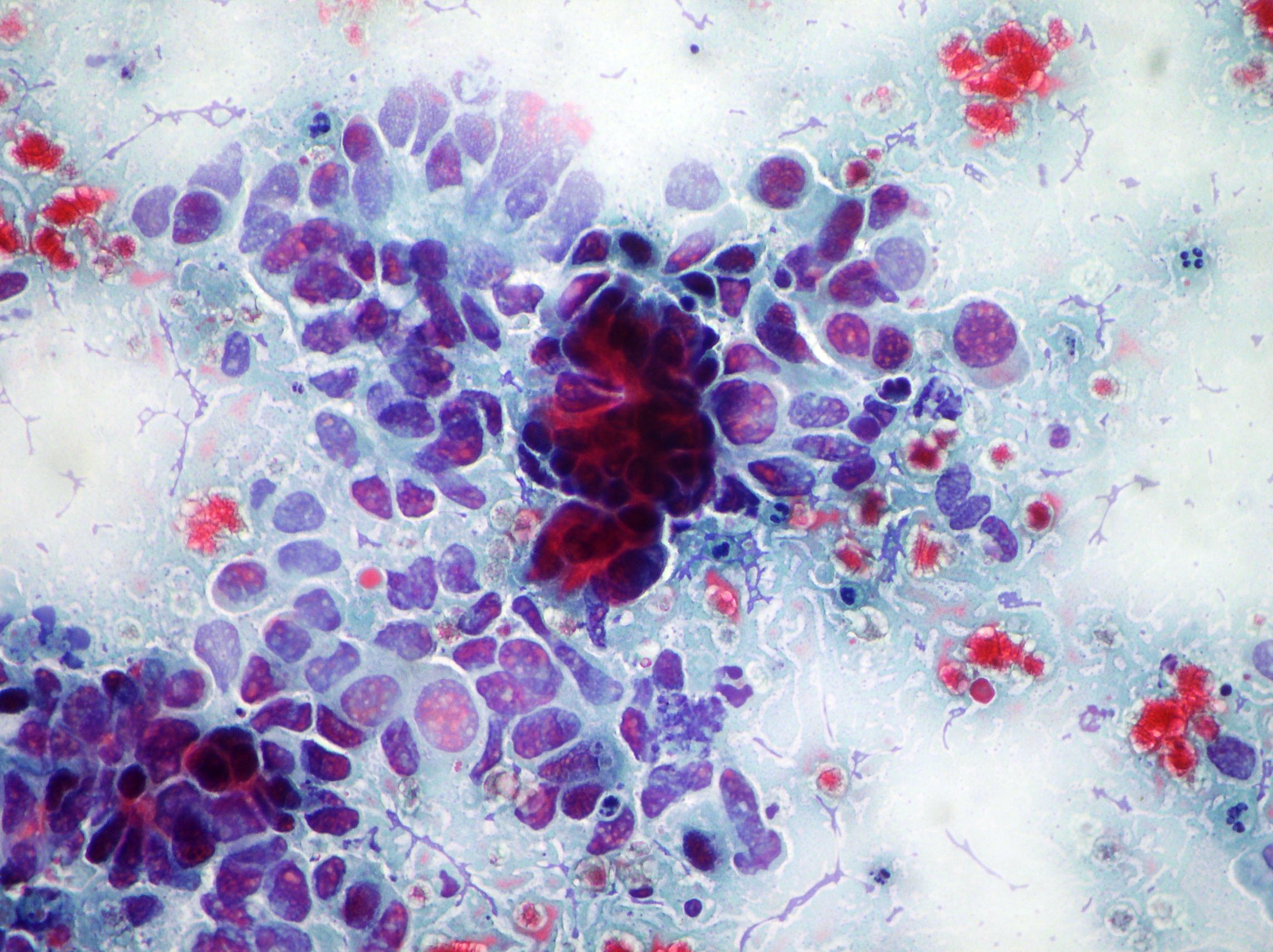
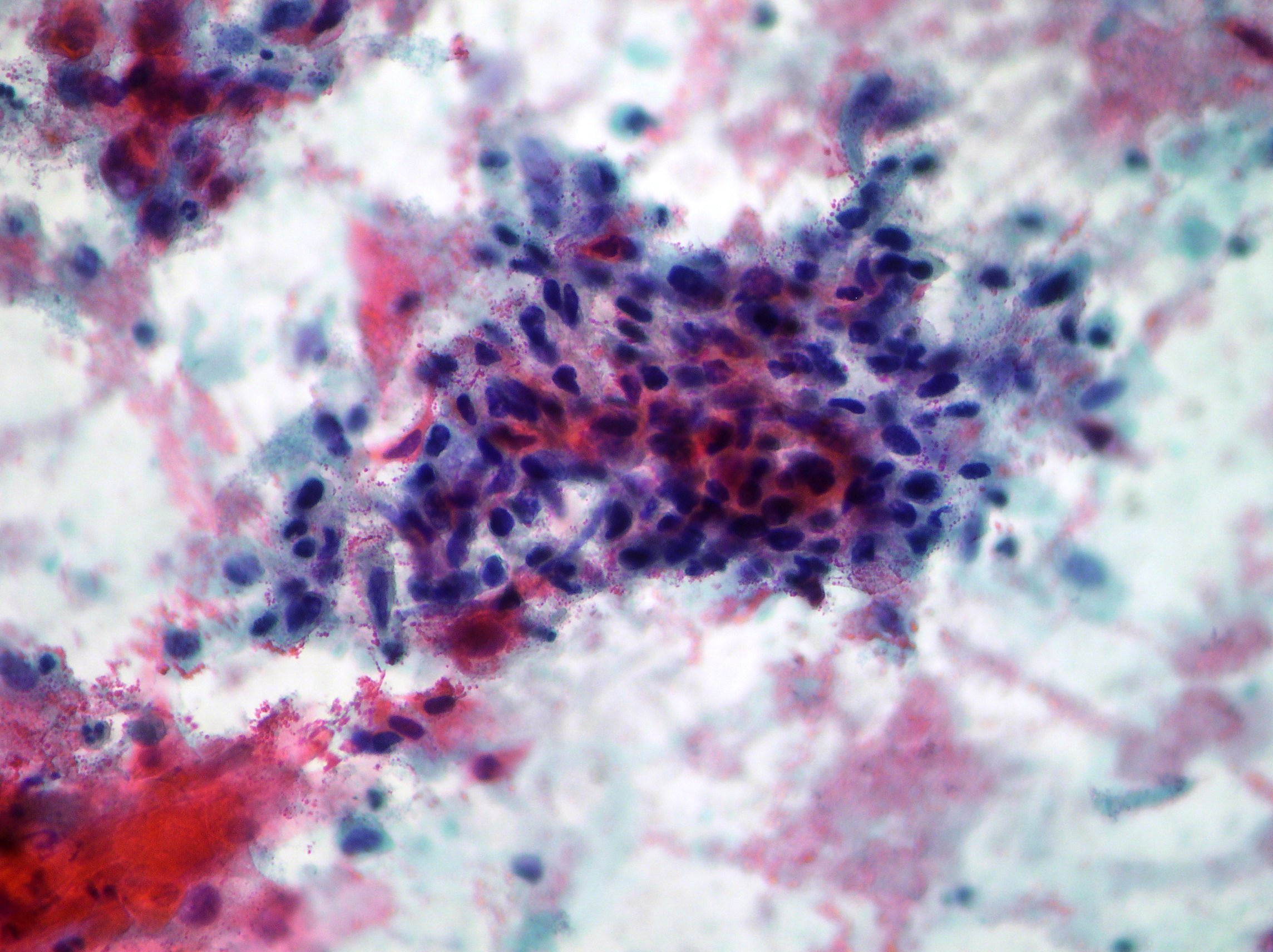
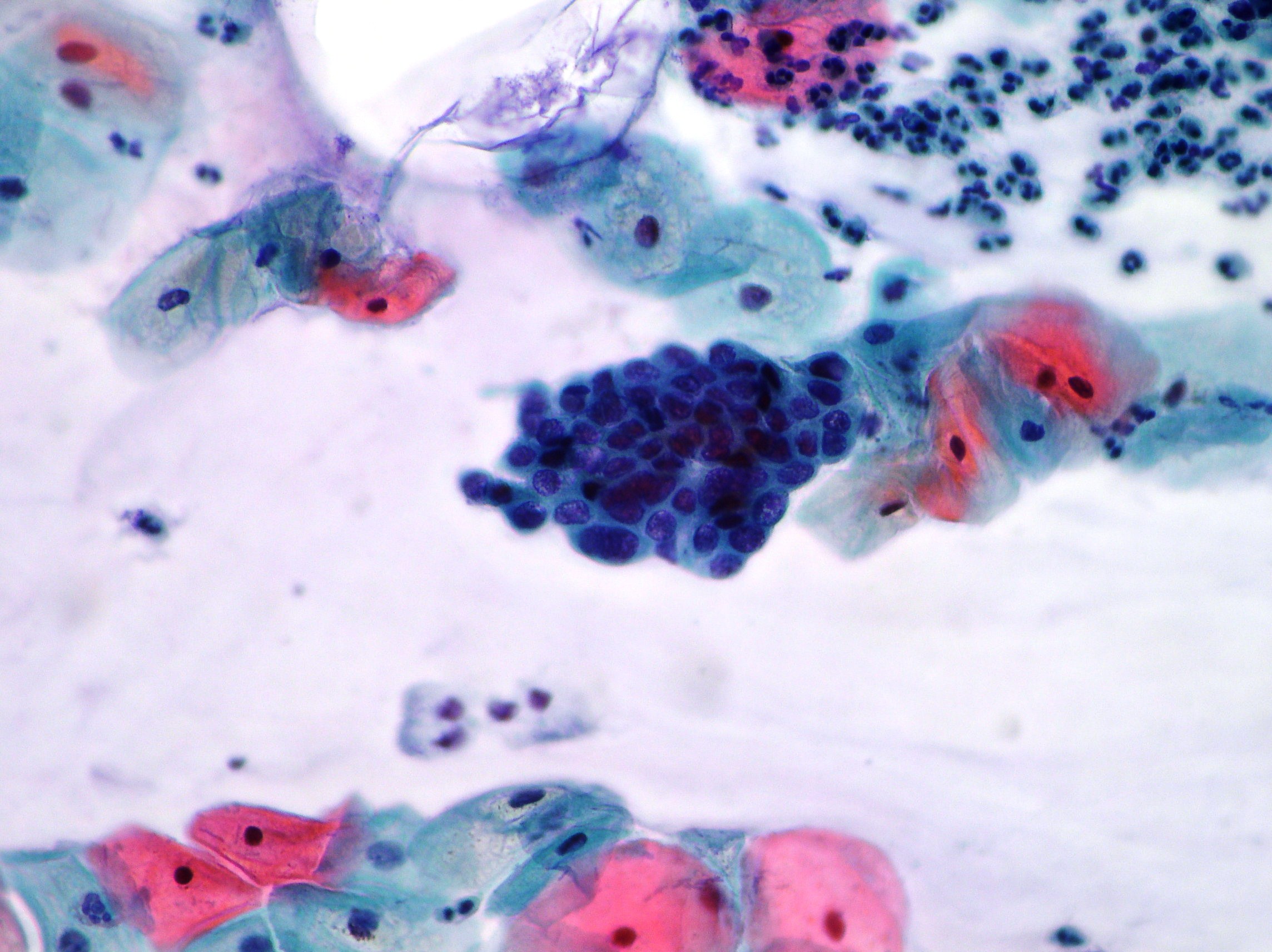
AGC
Abnormal glandular cells showing “fathering” and nuclear pseudostratification (Papanicolaou x100)
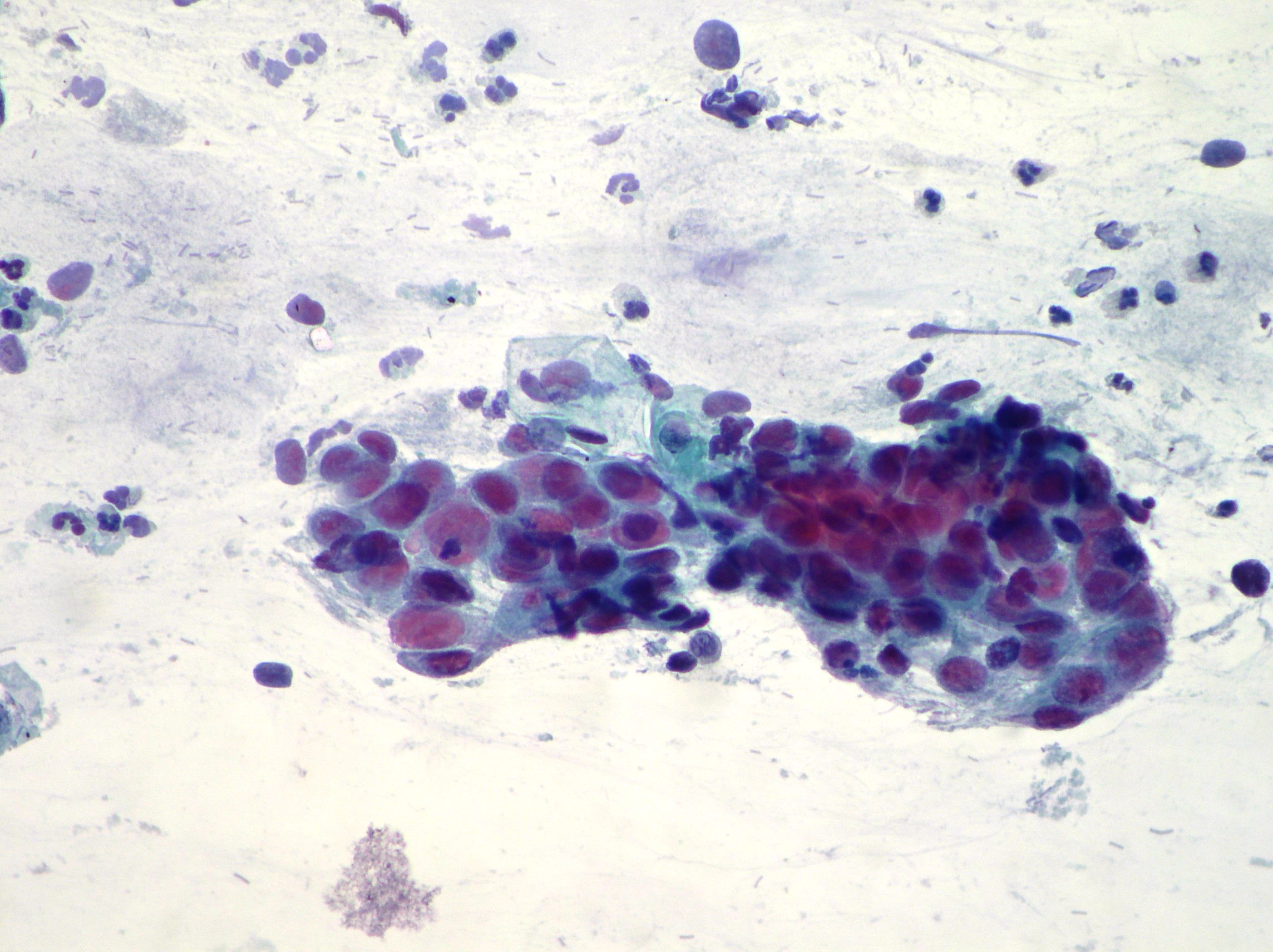
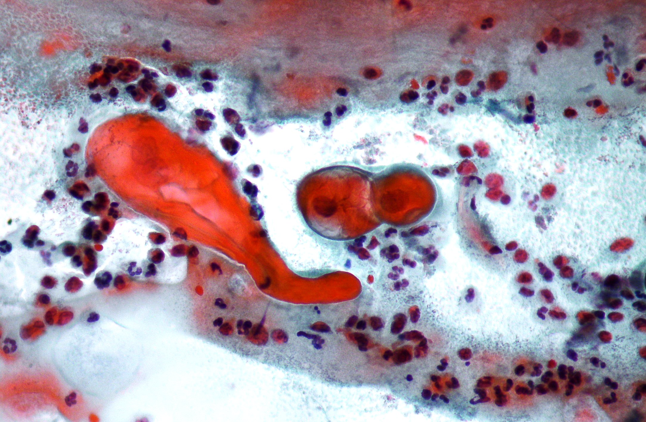
Squamous carcinoma of the uterine cervix
A case of keratinized squamous cell carcinoma of the uterine cervix. (Papanicolaou, x200) 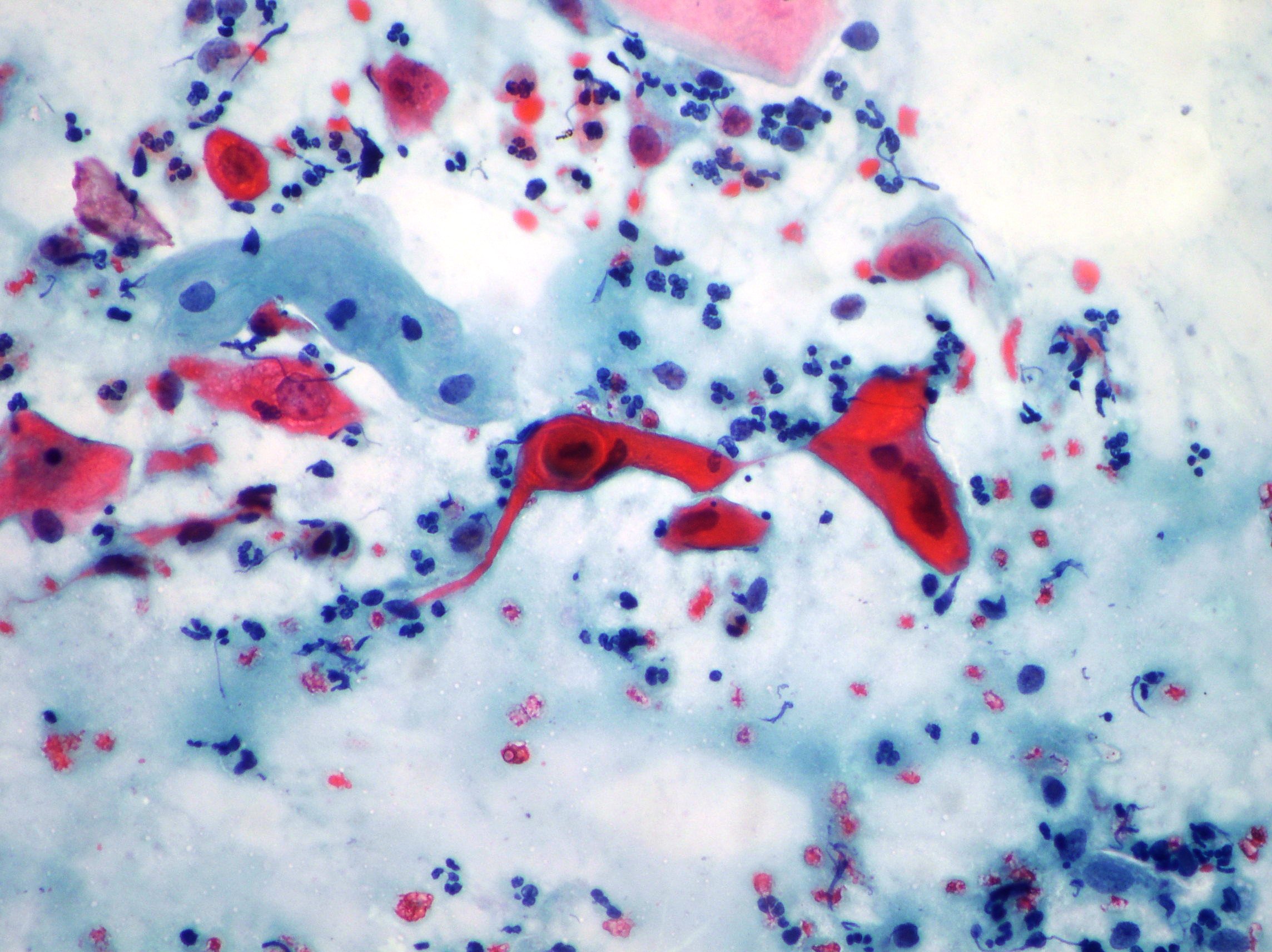
ASCUS-Atypical squamous cells of undetermined significance
Pap smear. Metaplastic cells sometimes binucleated and inflammation on the background suggestive of ASCUS. (Papanicolaou, x100) 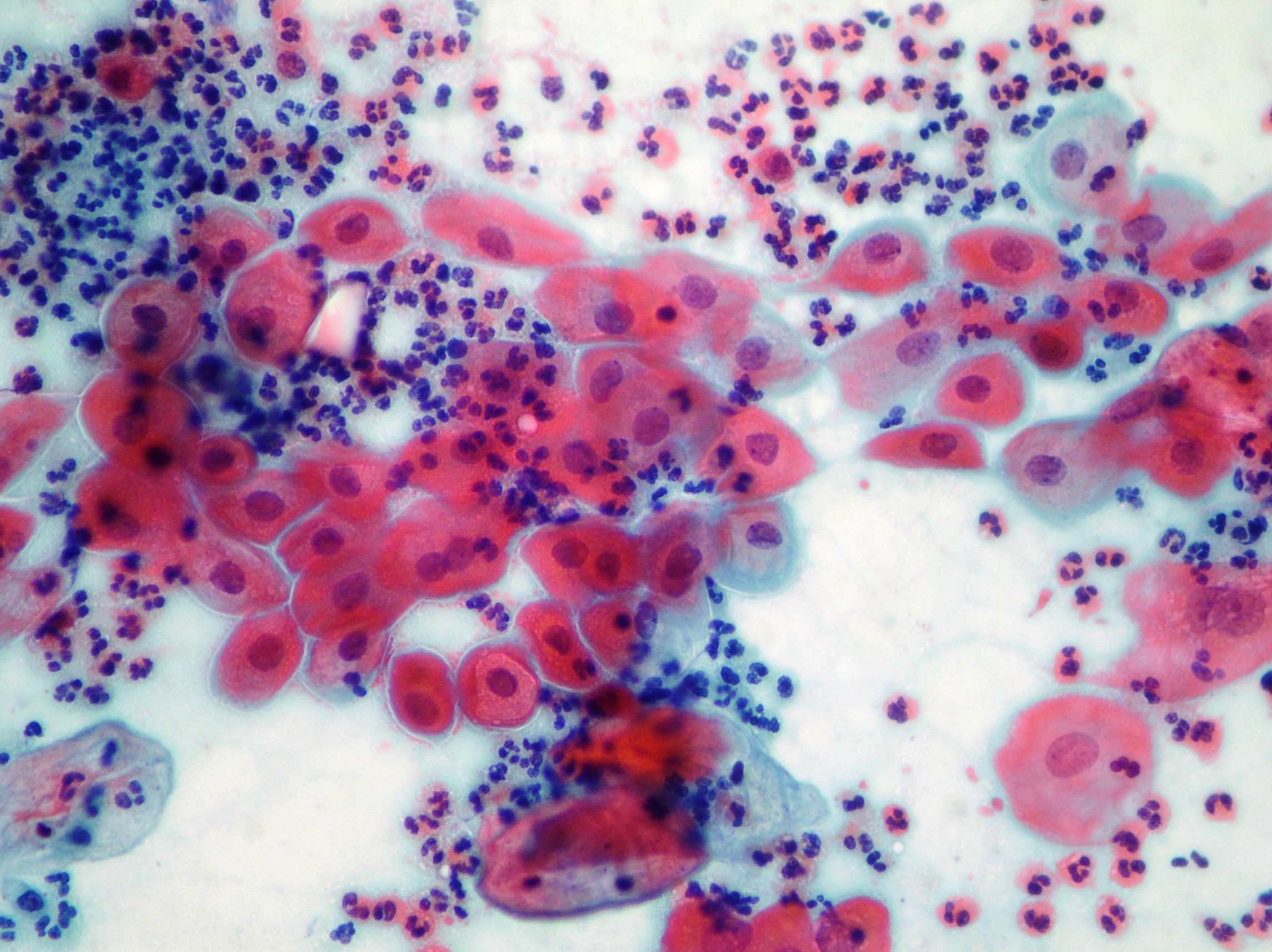
HSIL- High-grade squamous intraepithelial lesion
Epithelial cells showing moderate/severe nuclear alterations (Papanicolaou x100)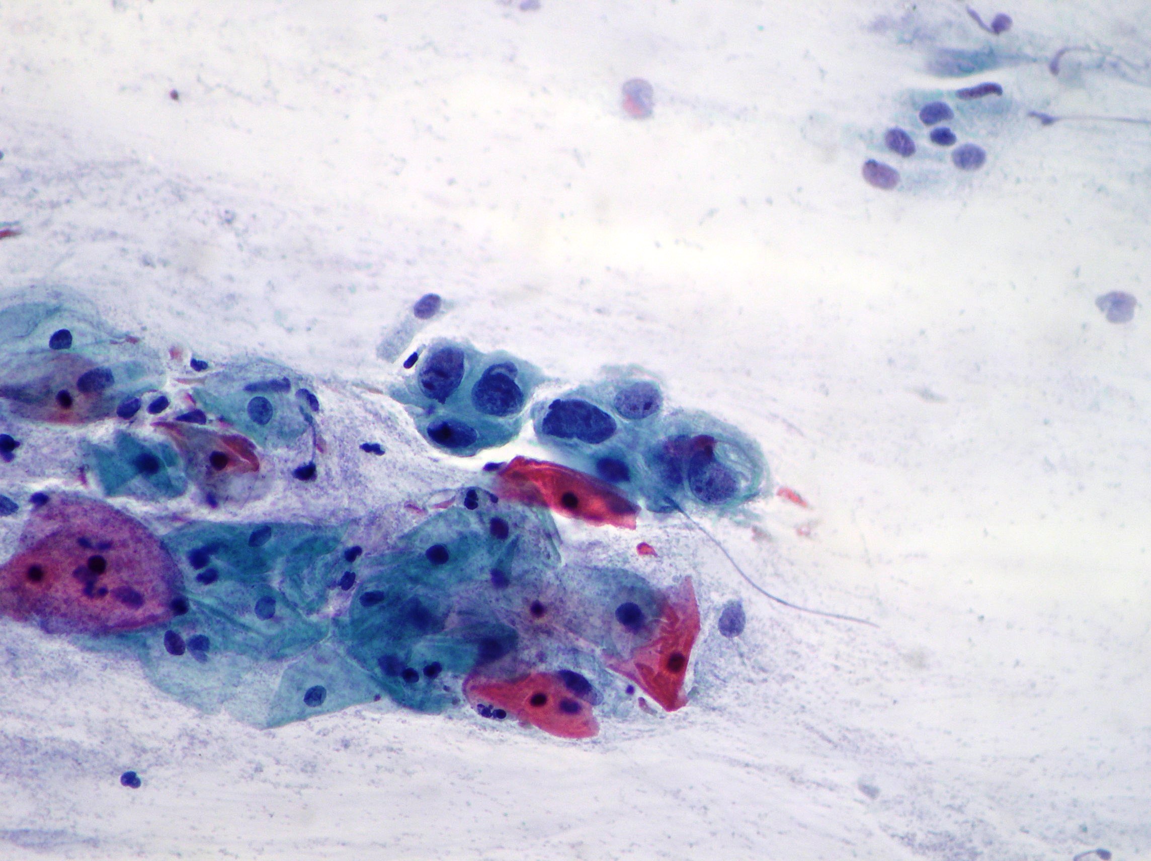
HSIL
HSIL with involvment of endocervical channel. (Papanicolaou x200)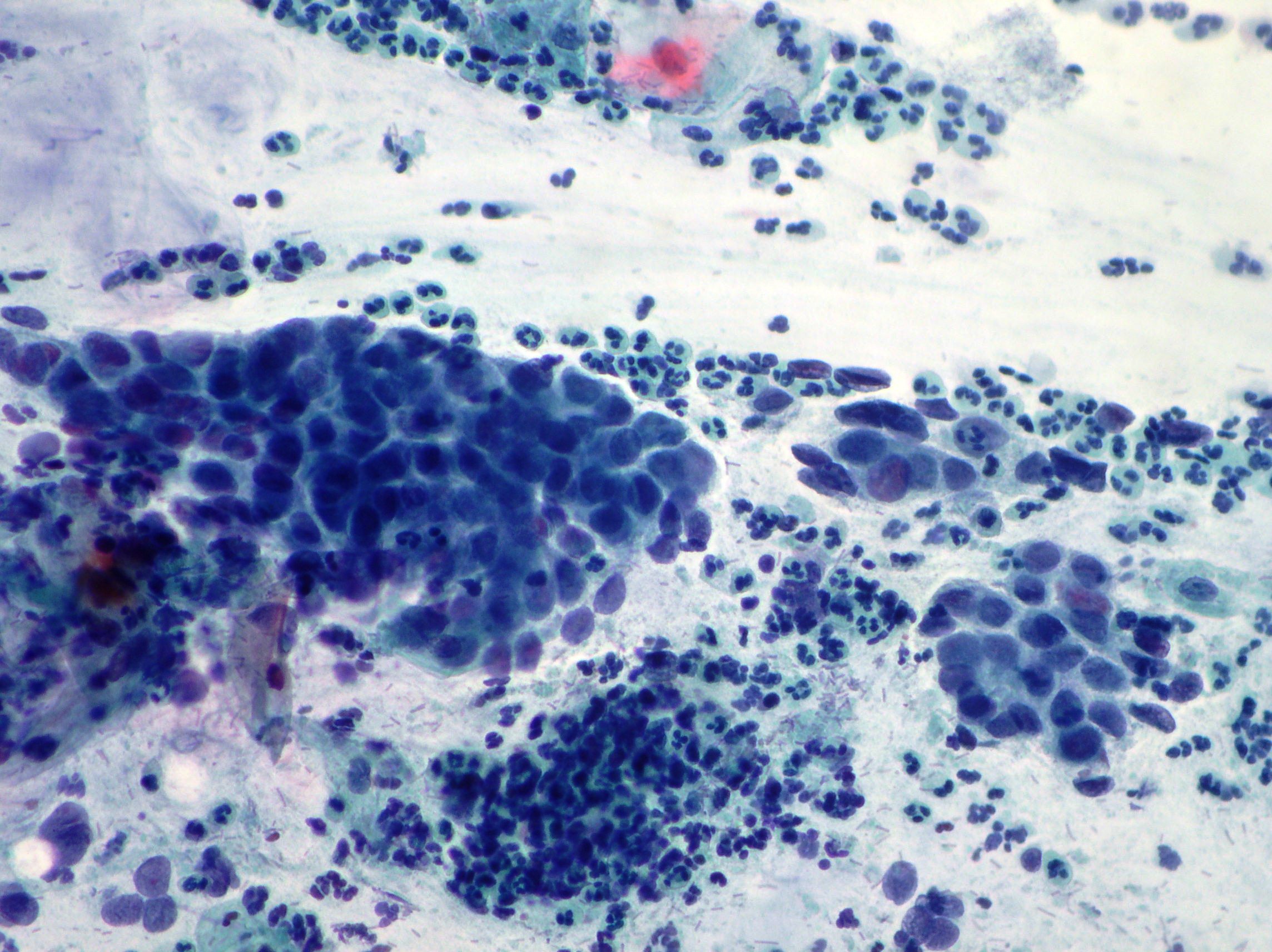
ASCUS
Atypical squamous cells of undetermined significance. (Papanicolaou x100)
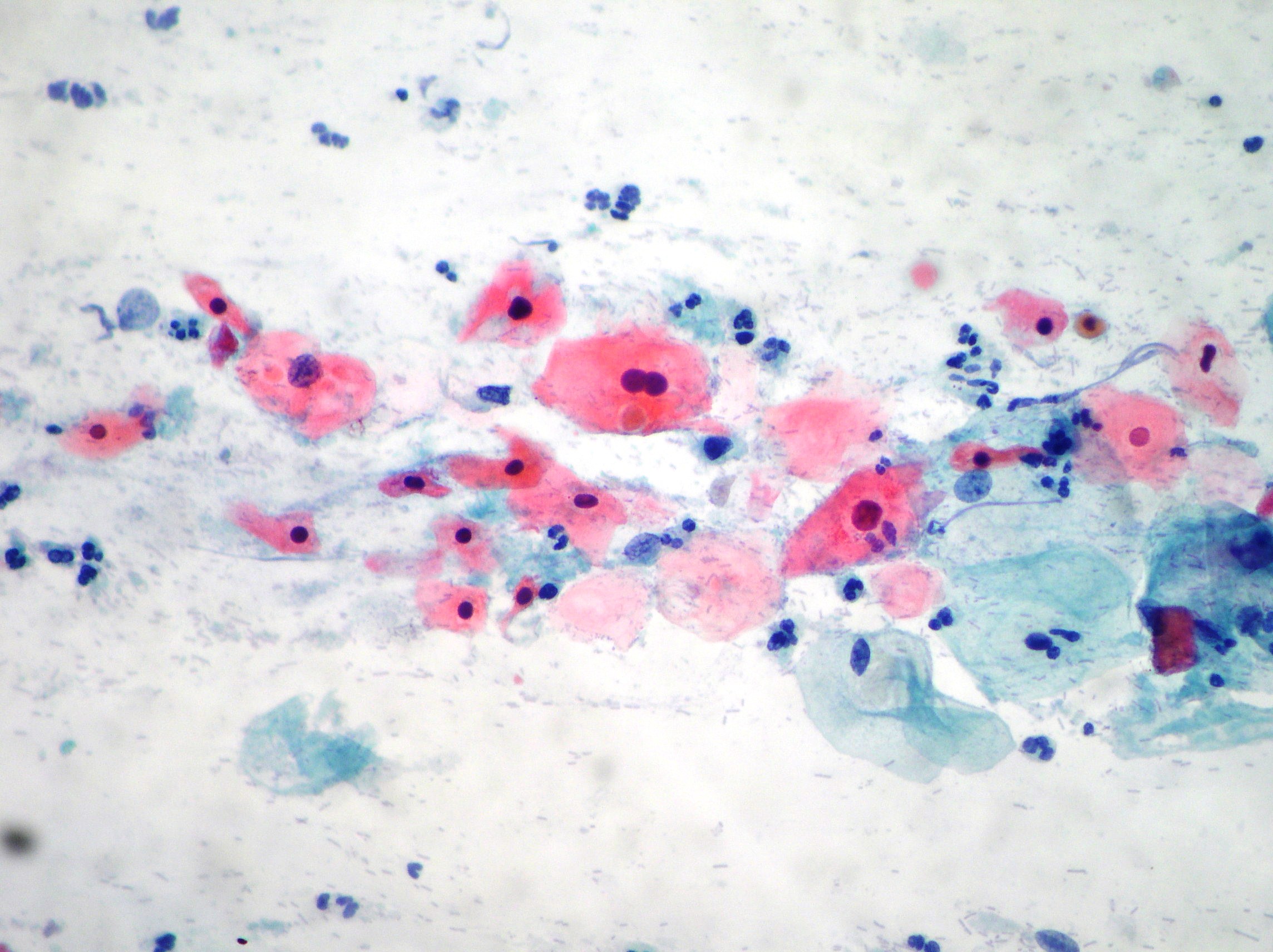
Squamous metaplasia
Mature and immature squamous metaplasia showing cells organized singly and on a sheet with relatively large nuclei and vacuolated cytoplasms. (Papanicolaou, x200)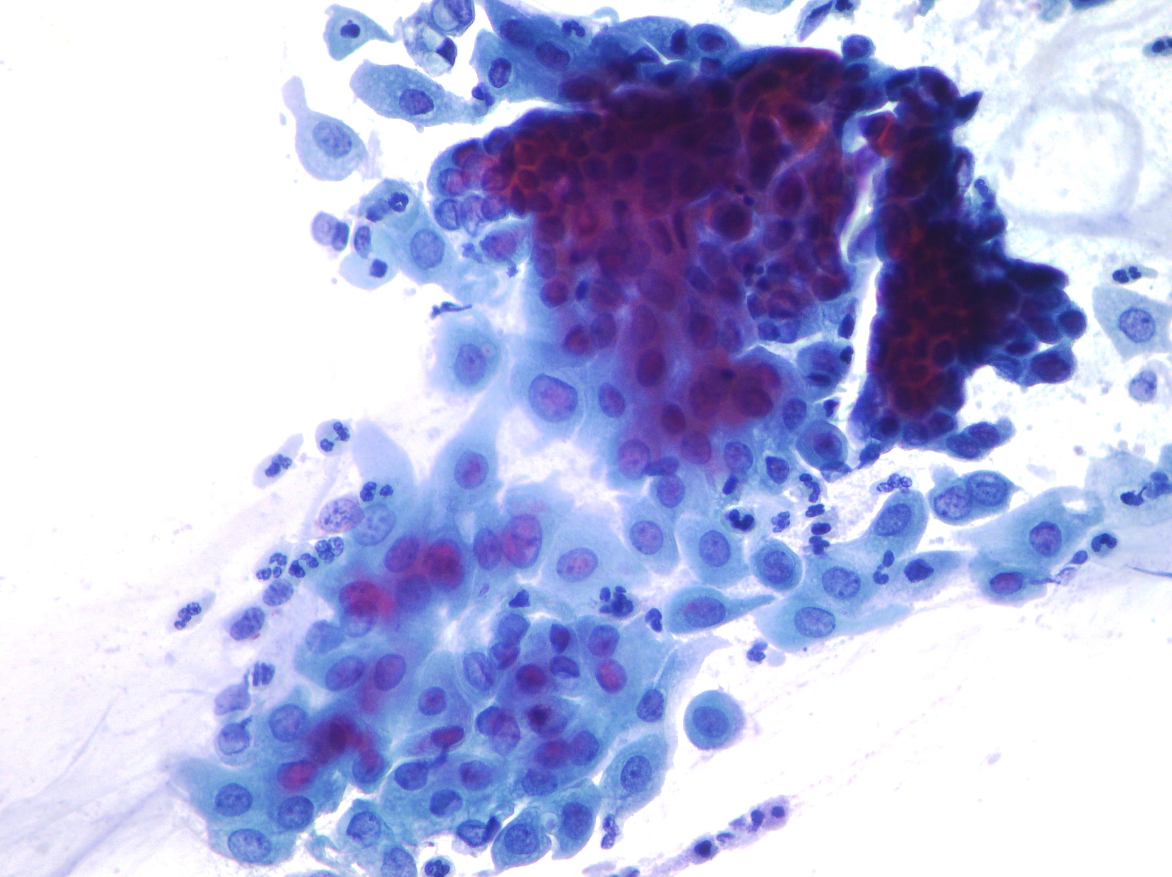
Keratinizing squamous cell carcinoma
Keratinizing squamous cell carcinoma showing bizzarre shaped cytoplasms and hyperchromatic nuclei. (Papanicolaou, x200)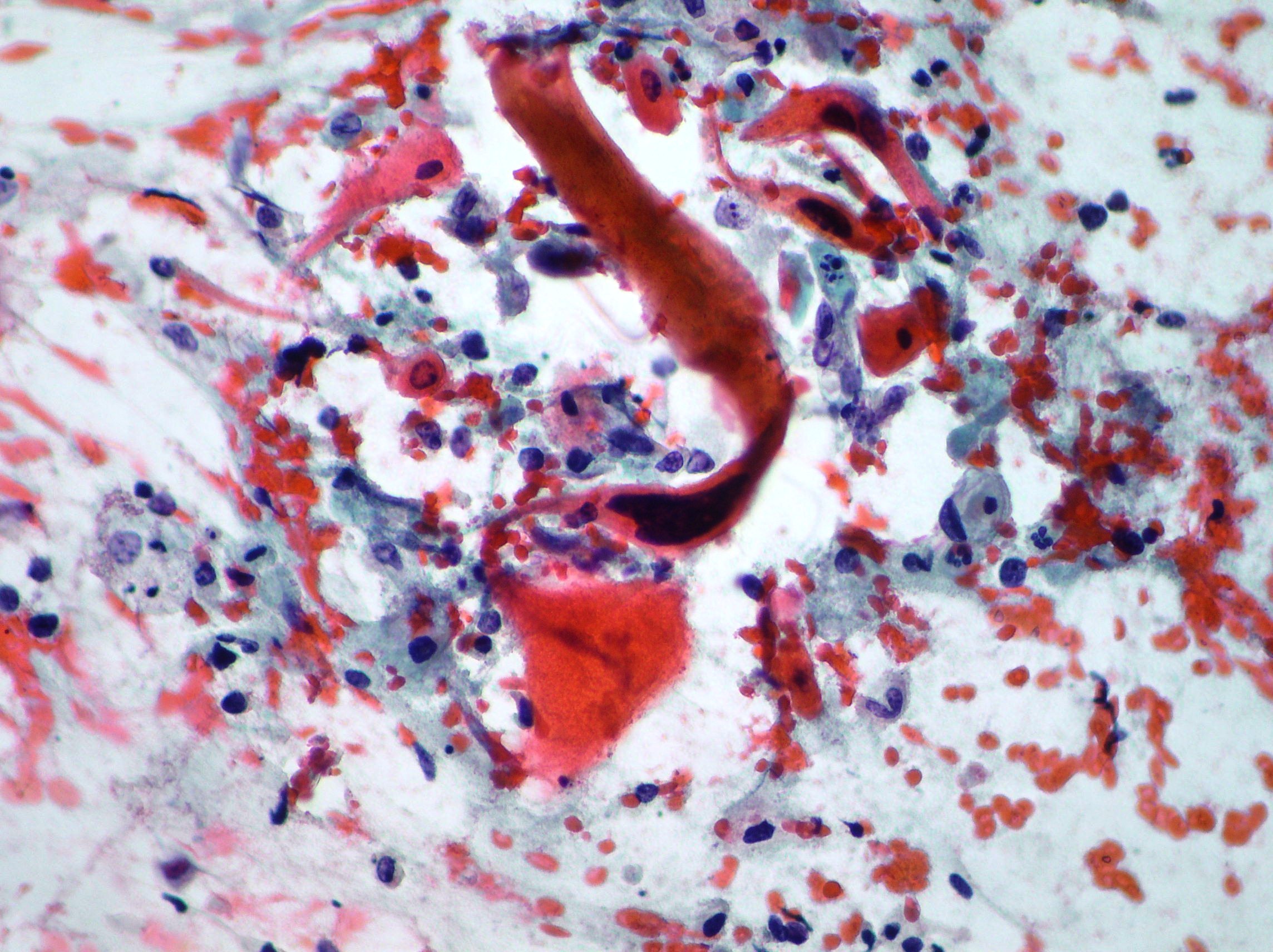
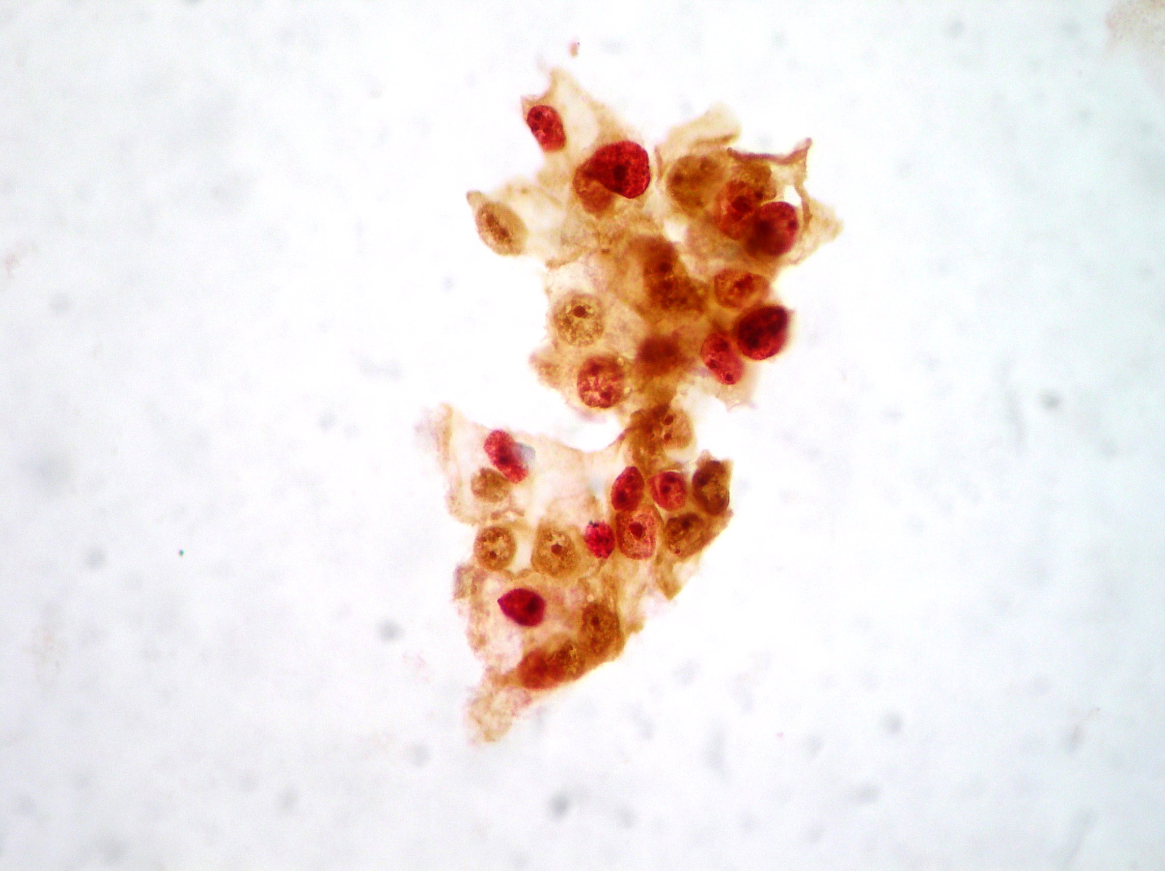
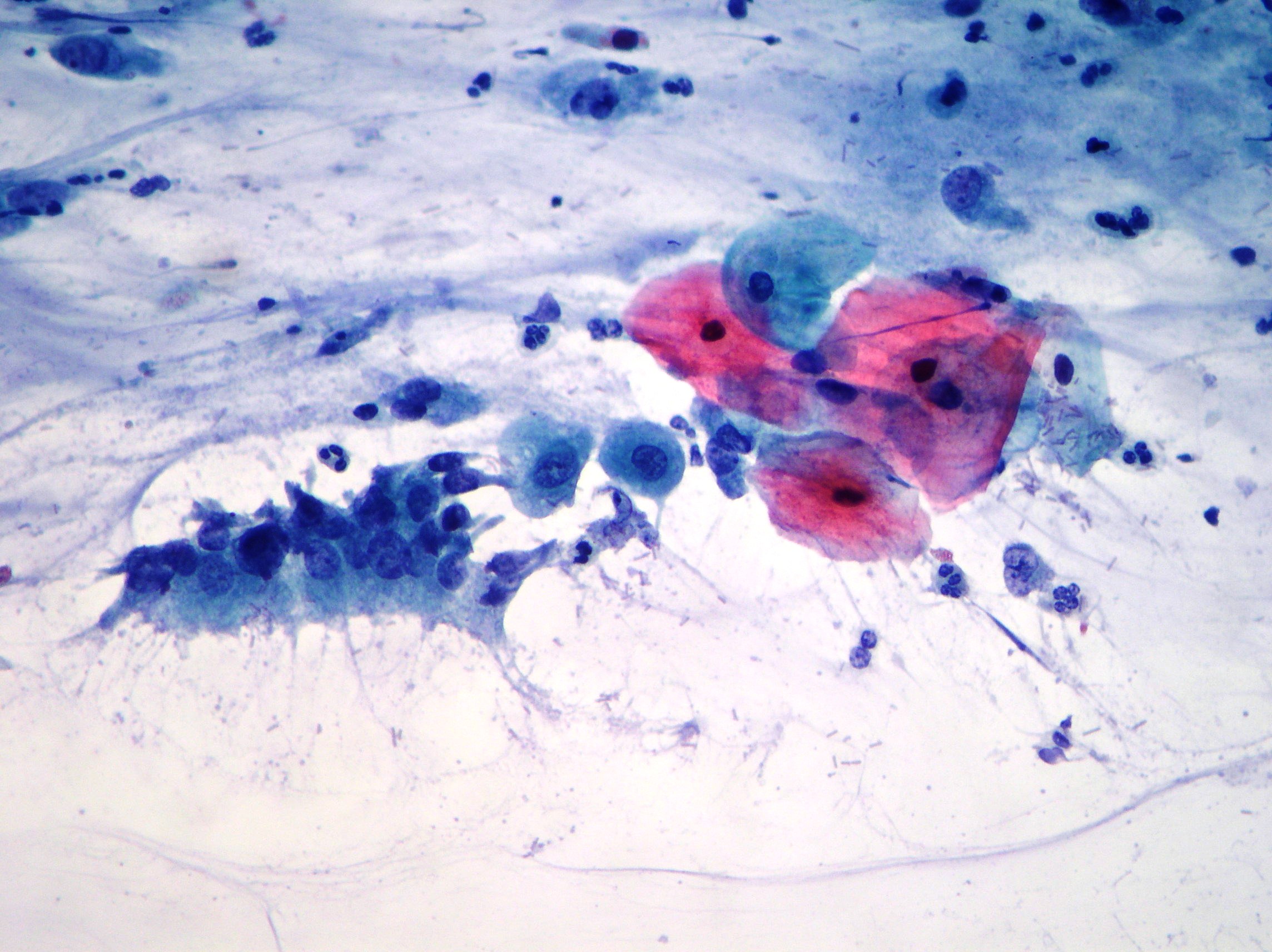
Radiation changes
Uterine cervix radiation changes exhibiting enlarged multinucleate cells with bizzarre shapes, vacuolated cytoplasms and phagocitosis of neutrophils. (Papanicolaou, 200)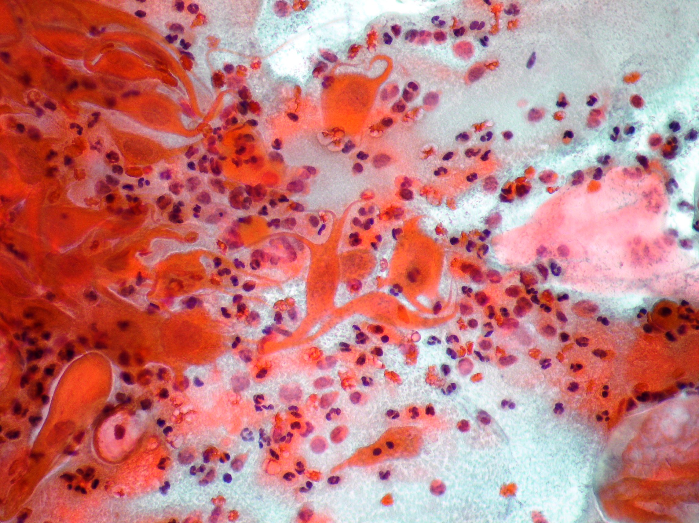
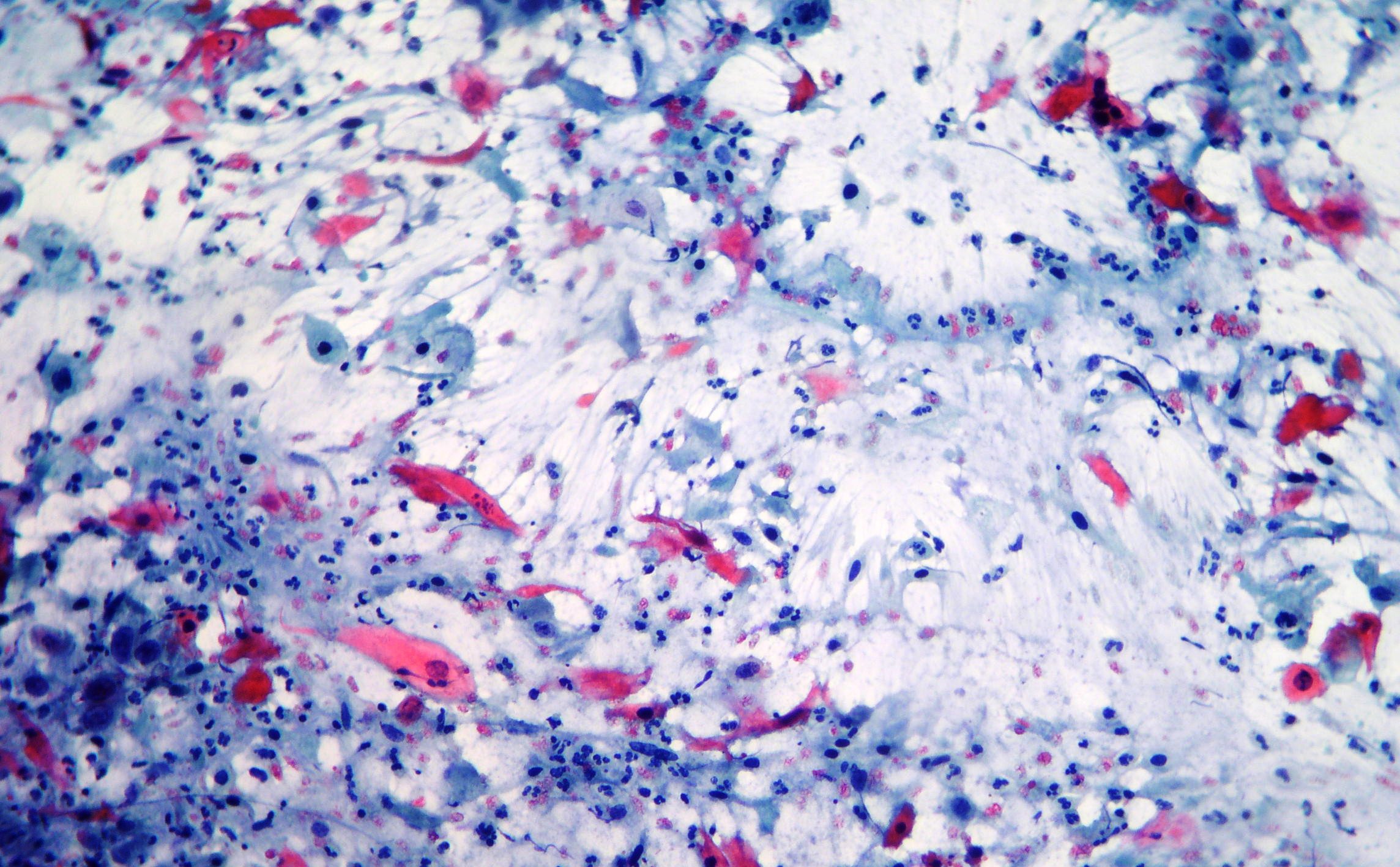
Keratinizing squamous cell carcinoma
Elongated and bizzarre shaped cells, degenerated round and oval nuclei with dense eosinophilic cytoplasm. Inflammation, debris and blood cells in the background. (Papanicolaou, x200, x100)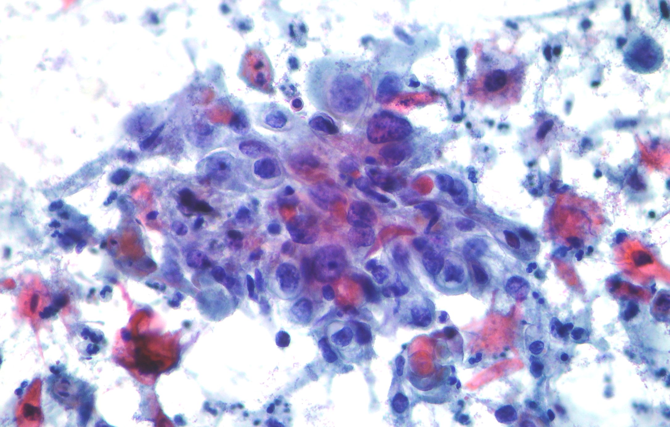
HPV infection effects
HPV infection show typical koilocytotic atypia exhibiting large perinuclear halos, condensation of the chromatin and slight atypia. (Papanicolaou, x600)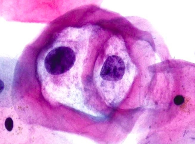
LSIL – Low grade squamous intraepithelial lesion
LSIL showing cells with vacuolization of the cytoplasm and slight atypia of the nuclei. (Papanicolaou, x400)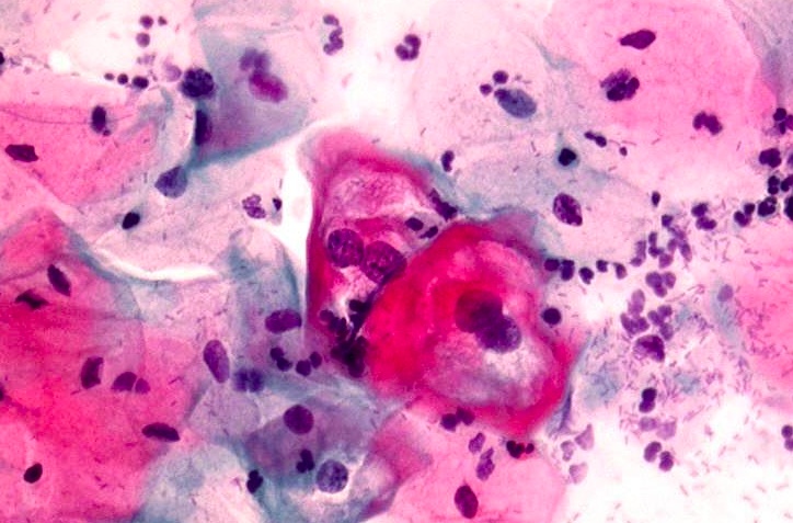
Normal pap smear
Normal superficial and intermediate squamous cell and tall columnar cells with eccentrically located nuclei. (Papanicolaou, x200)
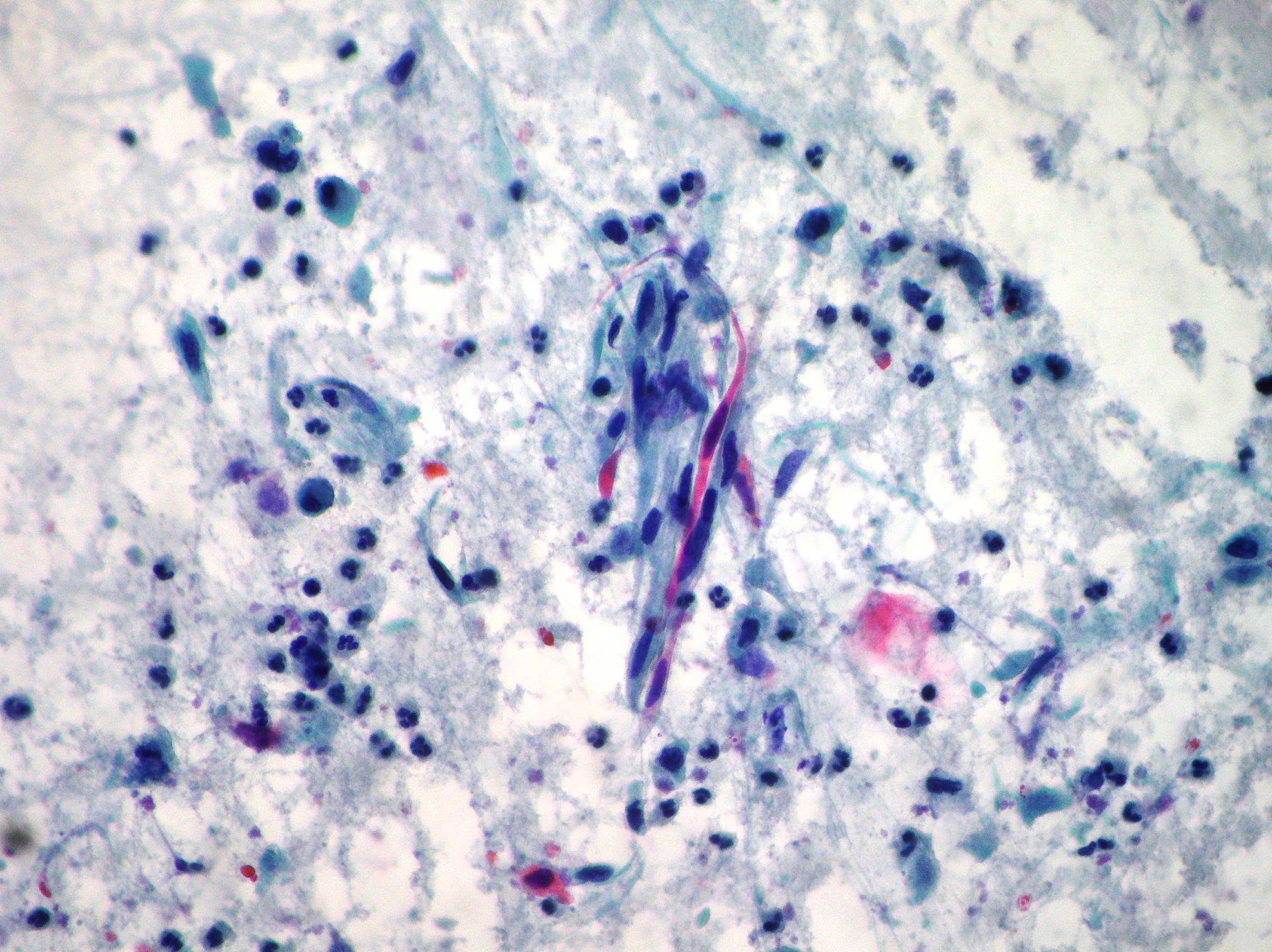
Herpes genitalis infection (HSV)
Herpes genitalis infection (HSV) showing atypical multinucleation, molding and small intranuclear chromatin granules suggestive of an early infection. (Papanicolaou, x200)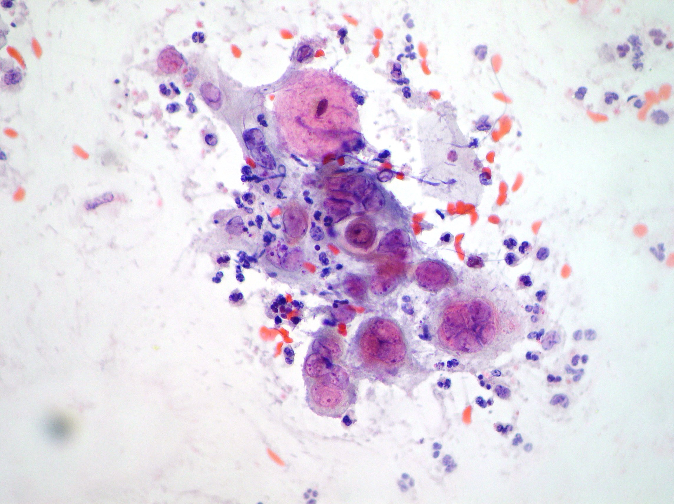
HSIL
Parabasal and basal cells showing hyperchromatic nucleus and high nucleocytoplasmic ratio suggestive of HSIL. (Papanicolaou, x200)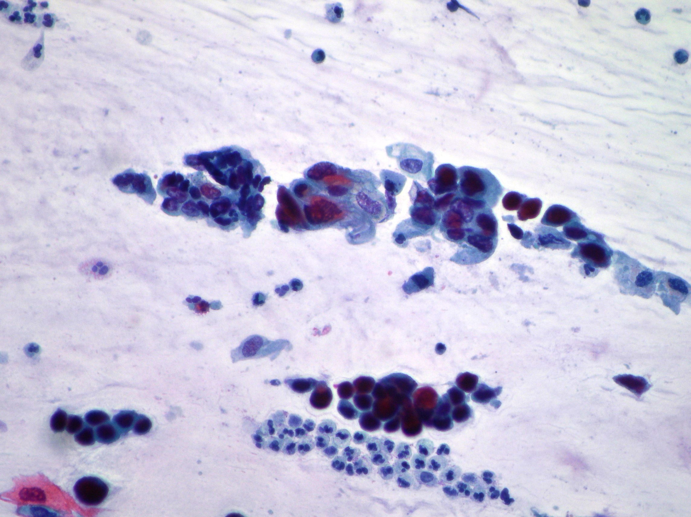
LSIL
HPV infection show typical koilocytotic atypia exhibiting perinuclear halos, condensation of the chromatin and binucleations. (Papanicolaou, x200)