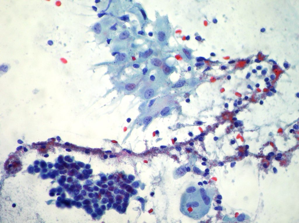
Reactive features.
Bronchial brushing. Normal bronchial cells associated to lymphocytes and epithelioid cells. (Papanicolaou x200)
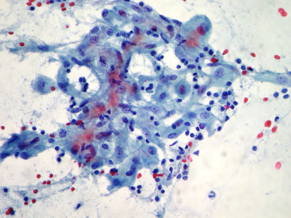
Normal and neoplastic cells.
Bronchial brushing. Normal and neoplastic cells in a case of squamous cell carcinoma. Papanicolaou x200
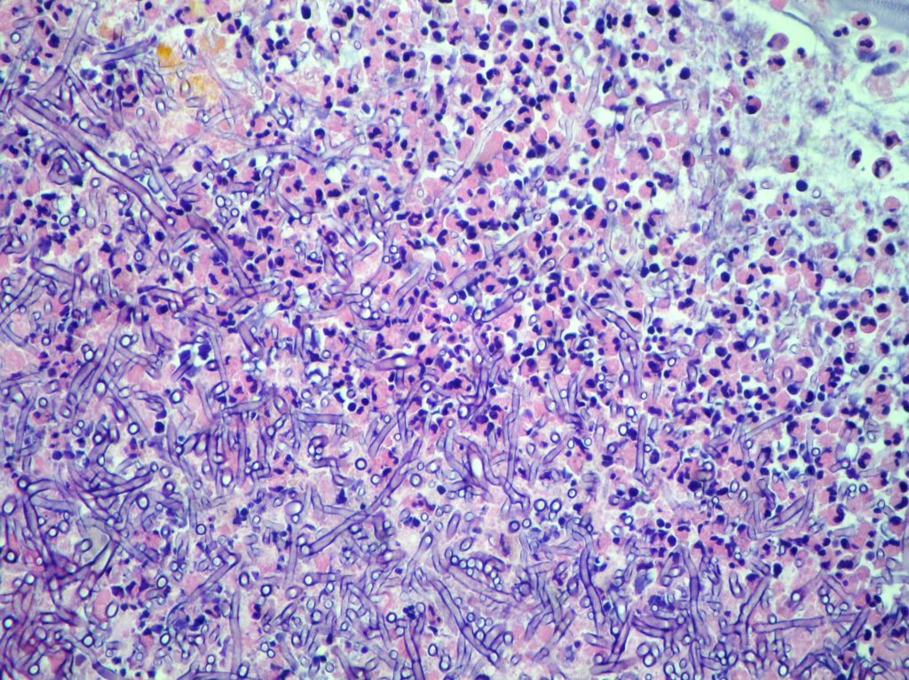
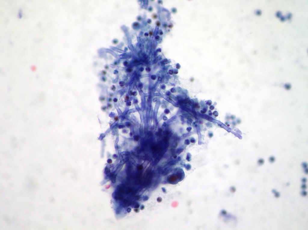
Pulmonary Mycetoma.
Bronchial washing. Necrotic material associated to inflammatory cells and several septet hyphae. CellBlock x100, Papanicolaou x200
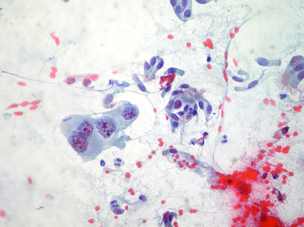
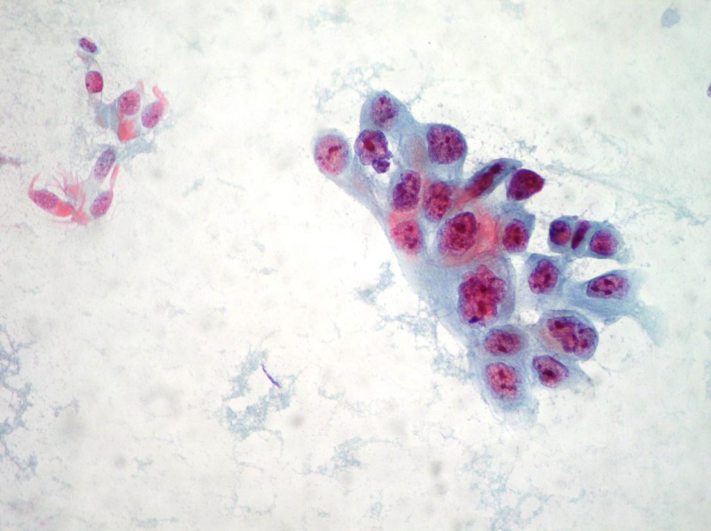
Bronchial washing.
Cluster of atypical cells (very suspected) arranged in papillary structures are resulted TTF1 (+) by immunocytochemistry and suggestive of adenocarcinoma. (Papanicolaou x200)
Lung squamous cell carcinoma.
Squamous cells carcinoma obtained from transthoracic needle aspiration. (Papanicolaou x200)
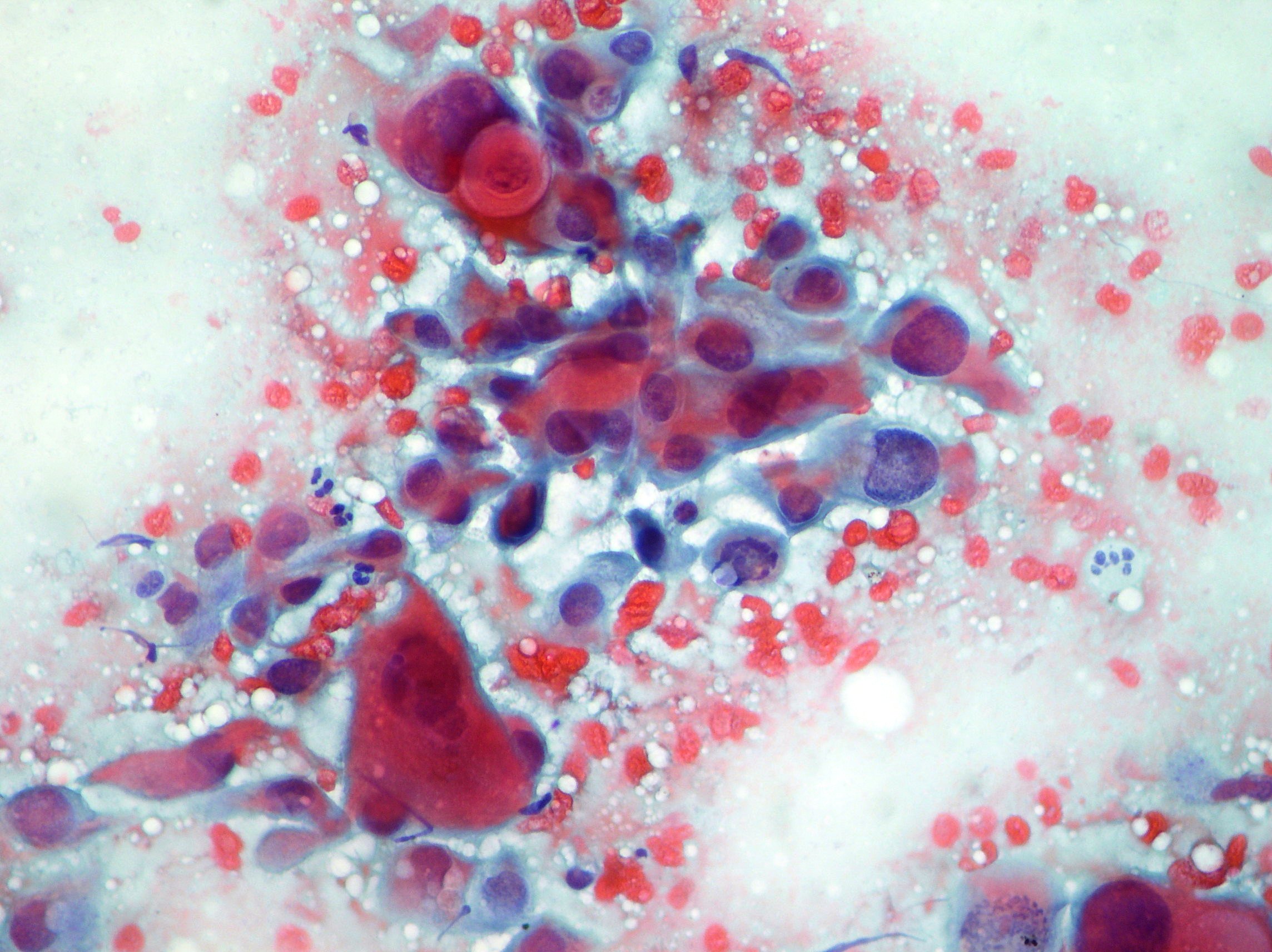
Lung Adenocarcinoma cells.
Adenocarcinoma cells obtained from transthoracic needle aspiration. (Papanicolaou x200; Diff Quick staining)
Squamous cell carcinoma.
Cancer cells obtained by bronchial brushing showing irregular clump of relatively cohesive pleomorphic cells. (Diff Quick staining x200; Papanicolaou x200)
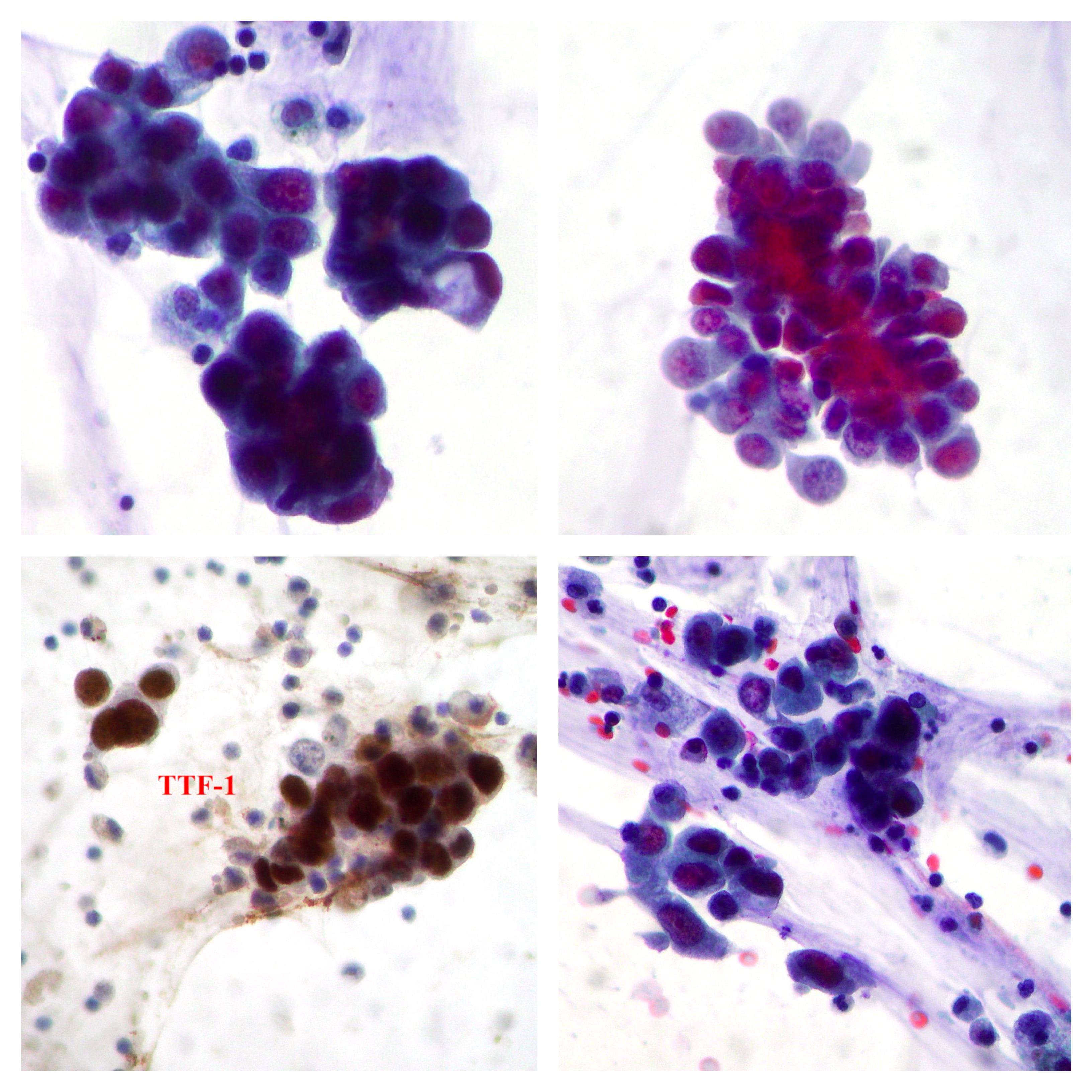
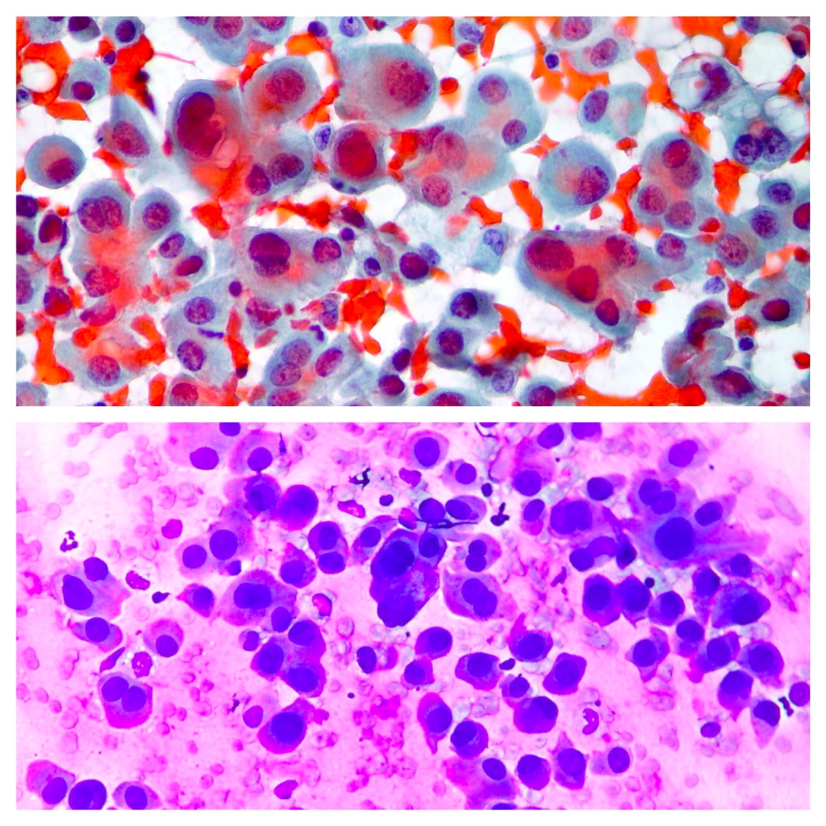
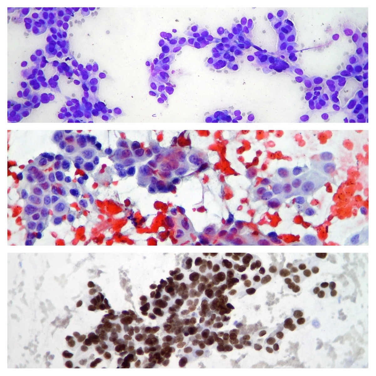
Adenocarcinoma cells.
Adenocarcinoma cells obtained by bronchial brushing. (Diff Quick staining x200; Papanicolaou x200; TTF1 immunocitochemistry staining)
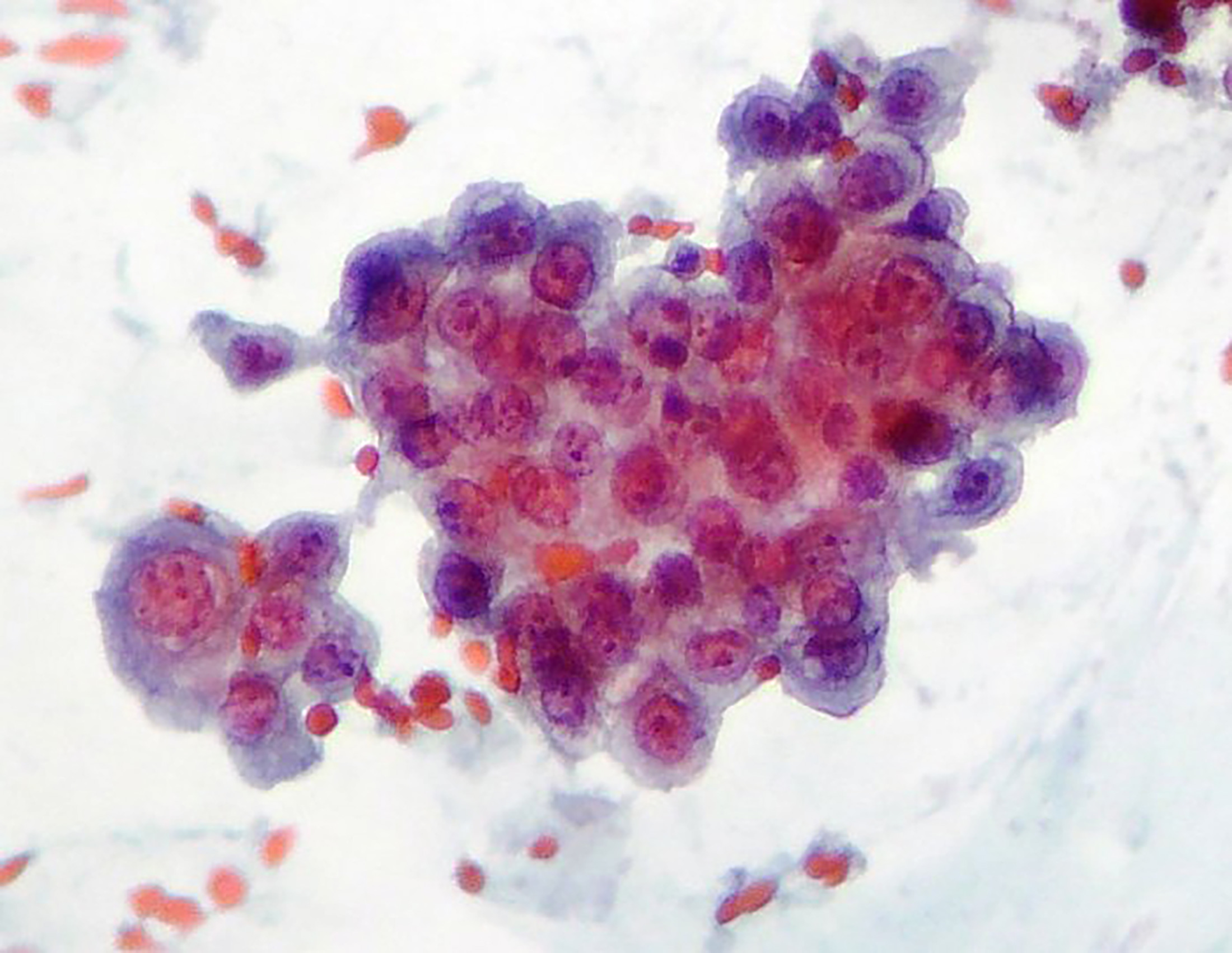
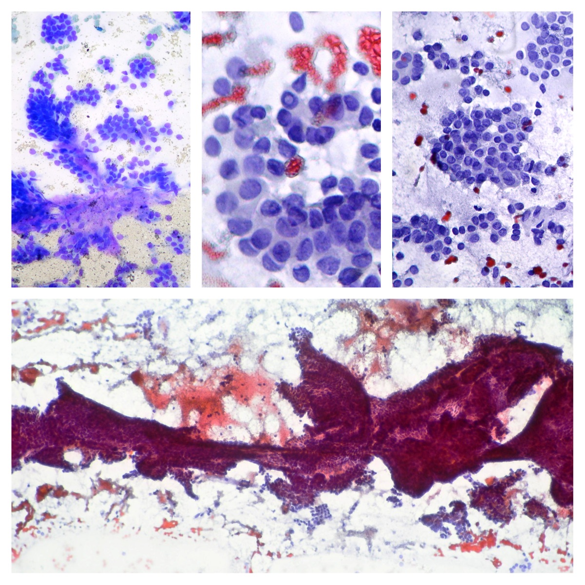
Lung metastatic of papillary thyroid carcinoma.
A case of metastatic papillary thyroid carcinoma obtained from transthoracic needle aspiration. Sheets of papillary carcinoma with oval, pale nuclei and intranuclear inclusion. (Diff Quick and Papanicolaou staining)
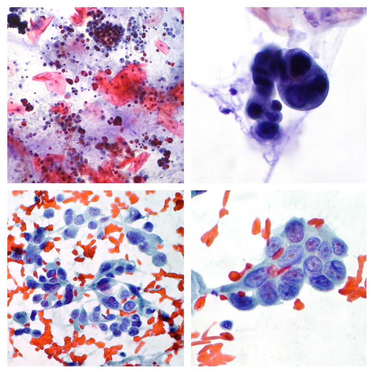
Adenocarcinoma cells
Sputum (upper side) and transthoracic needle aspiration from the same case. Cells are suggestive of adenocarcinoma. (Papanicolaou x100, x200)
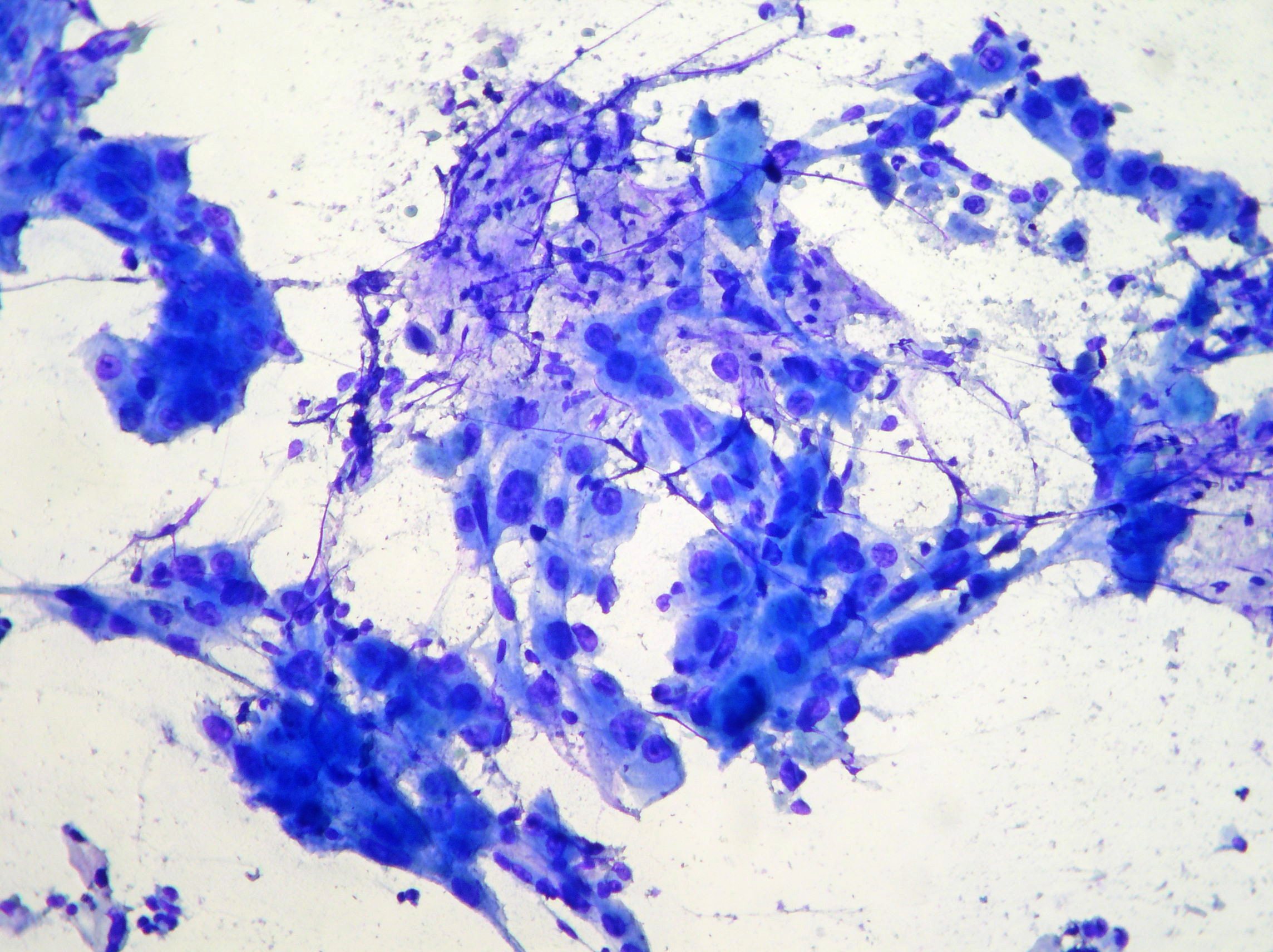
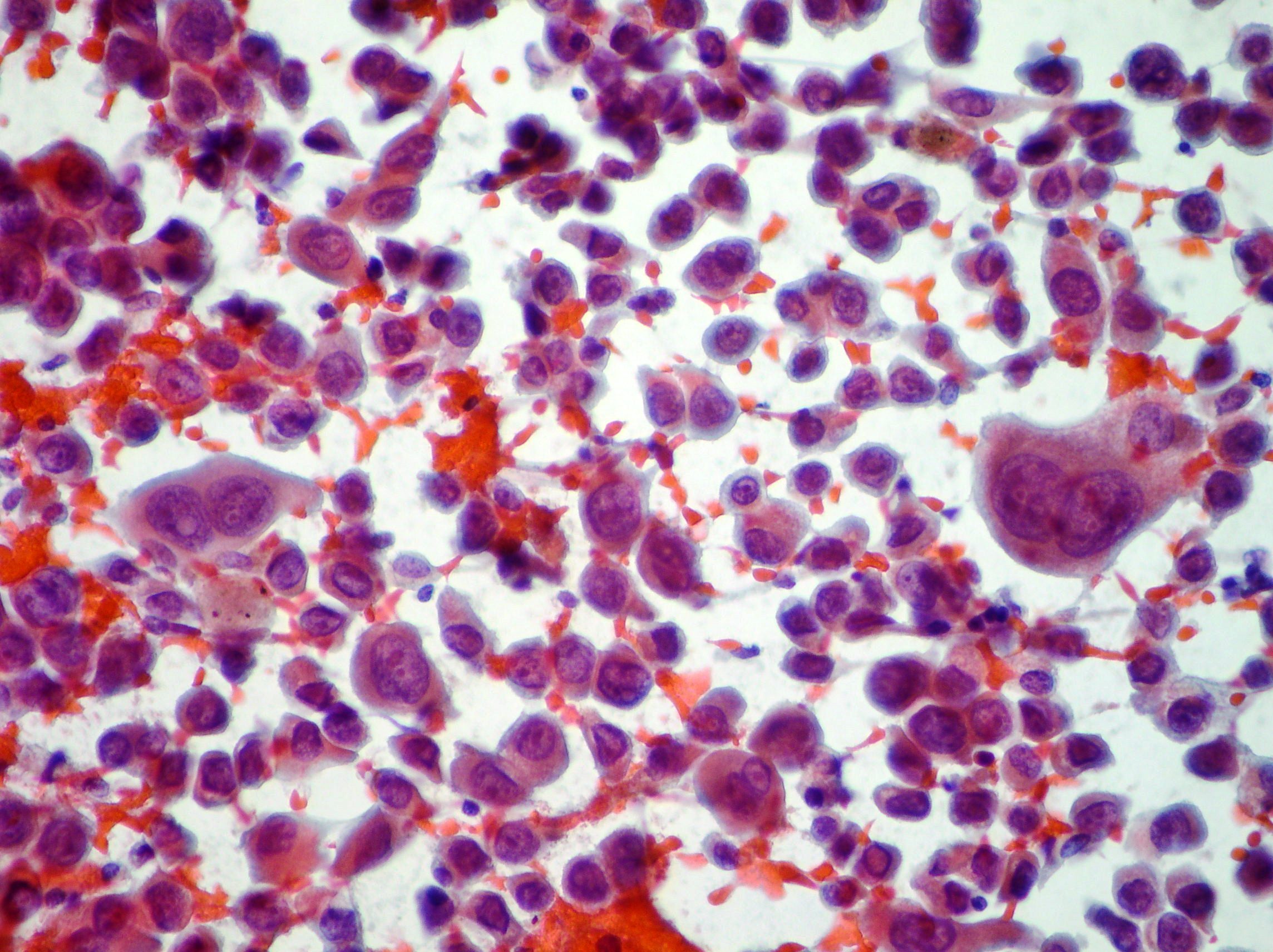
NSCLC
Smears of malignant cells suggestive of NSCLC. (Papanicolaou x200; Diff Quick staining)
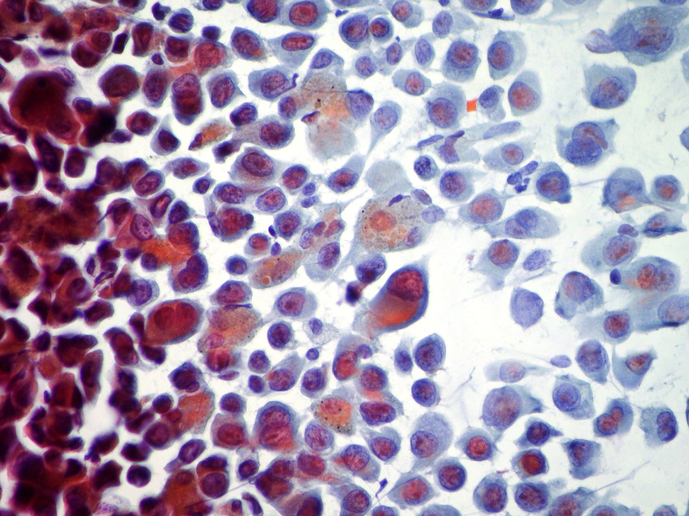
Undifferentiated carcinoma cells
Undifferentiated carcinoma cells showing prominent anisokariosis and macronucleoli. (Papanicolaou x400; Diff Quick staining)
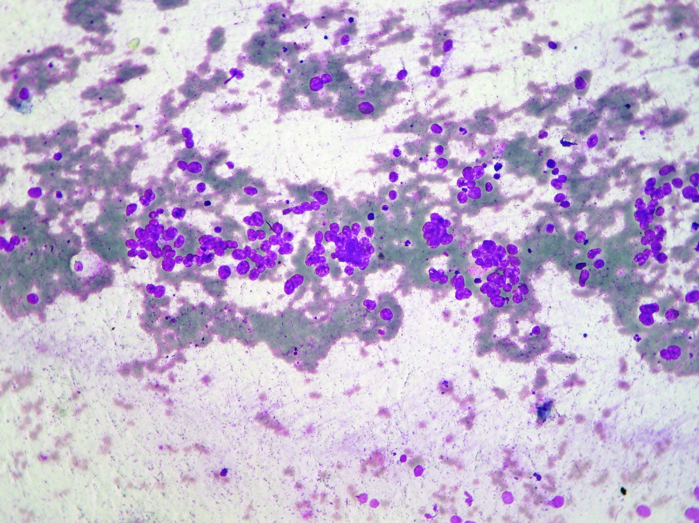
Lung adenocarcinoma cells.
Lung adenocarcinoma cells obtained from Transthoracic needle aspiration. (Diff Quick staining)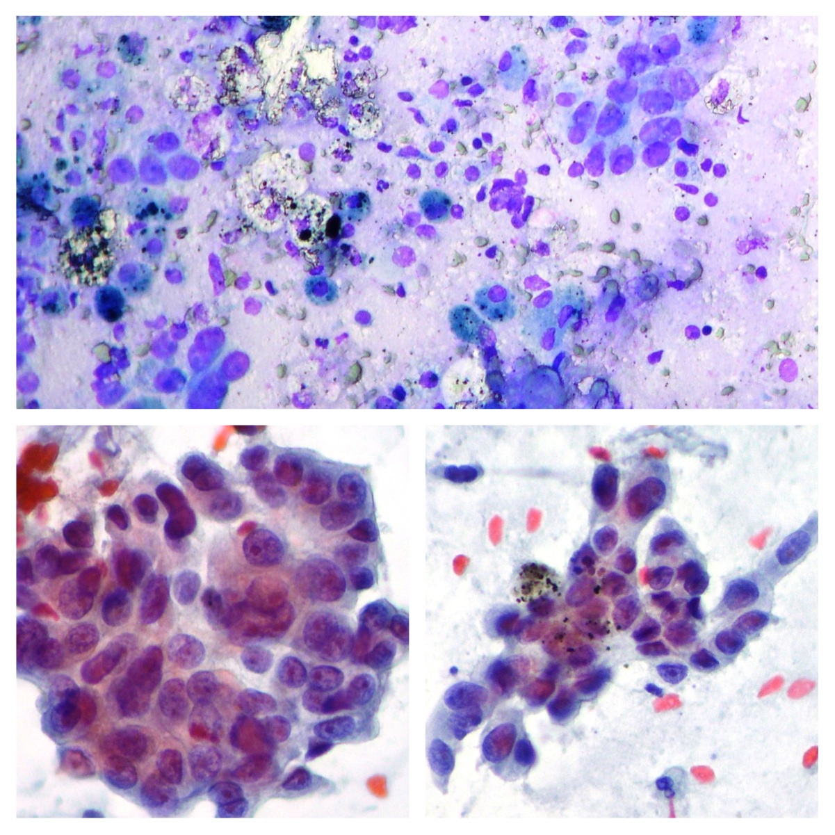
NSCLC
Carcinoma cells showing pleomorphic malignant cells and necrotic debris compatible with NSCLC. (Papanicolaou x400; Diff Quick staining)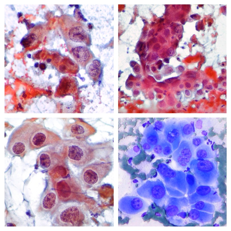
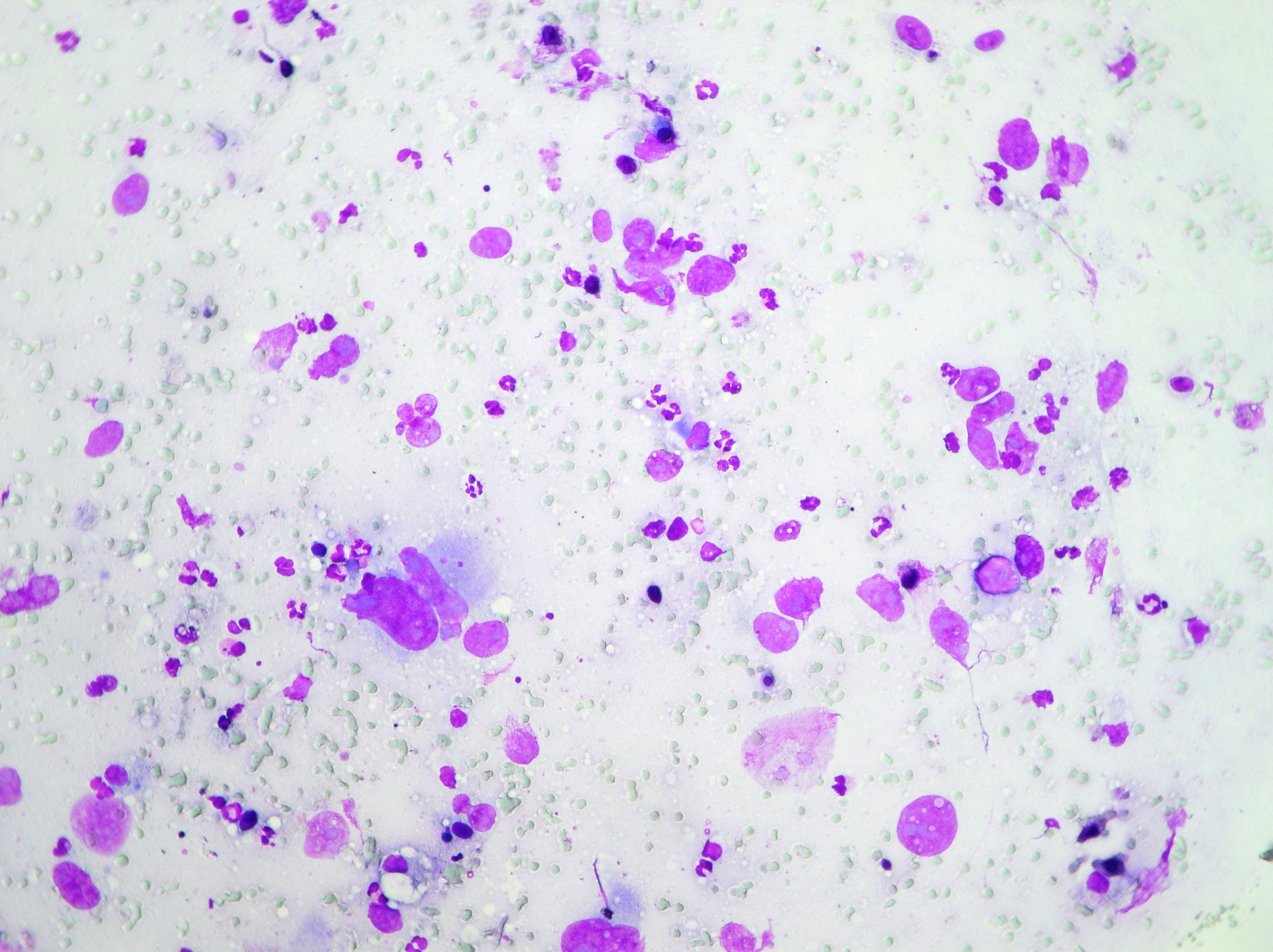
Small cell lung carcinoma
Smears of small cell lung carcinoma showing pleomorphism, poorly cohesive cells, nuclear molding. (Papanicolaou x200; Diff quick staining)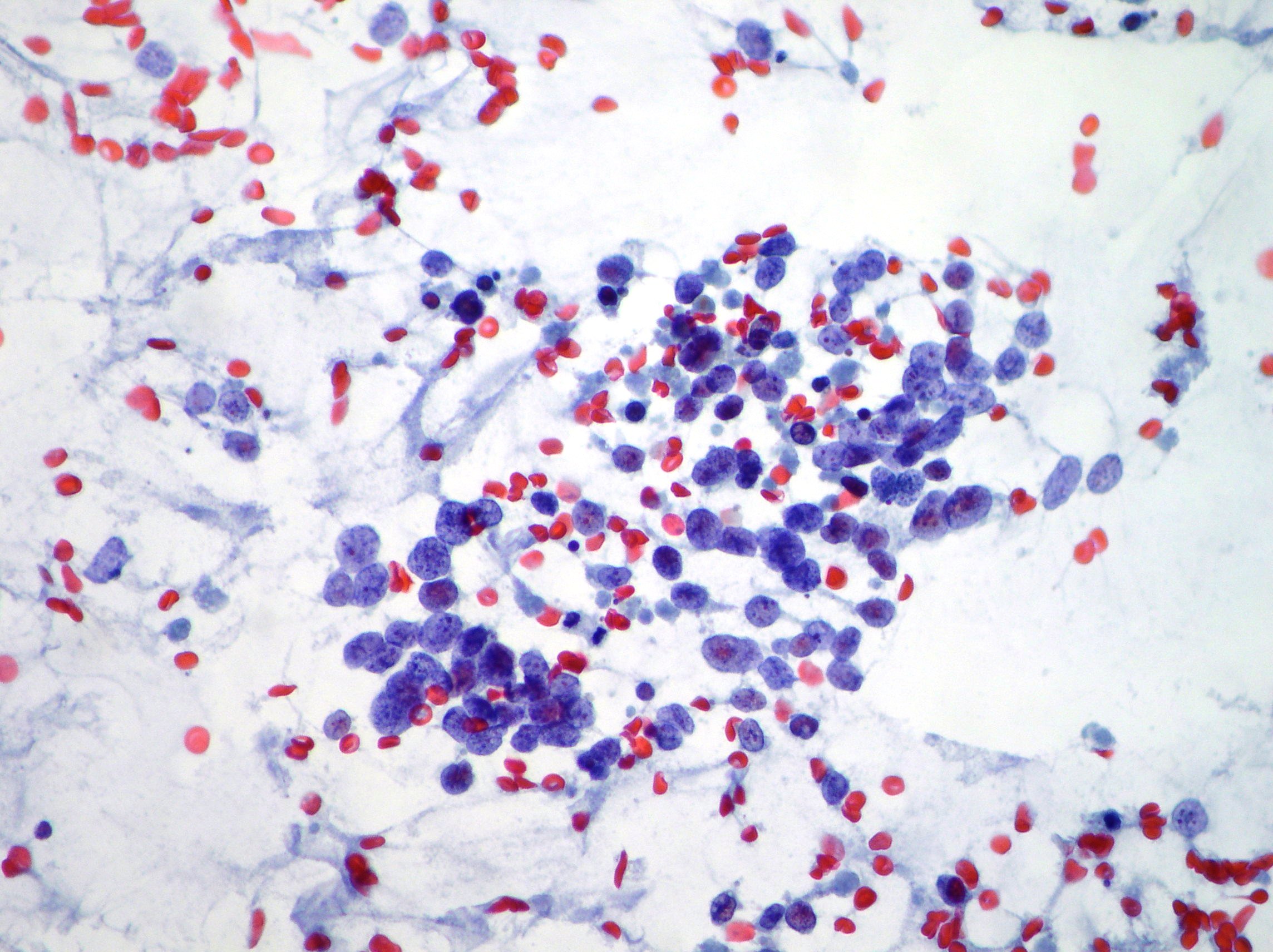
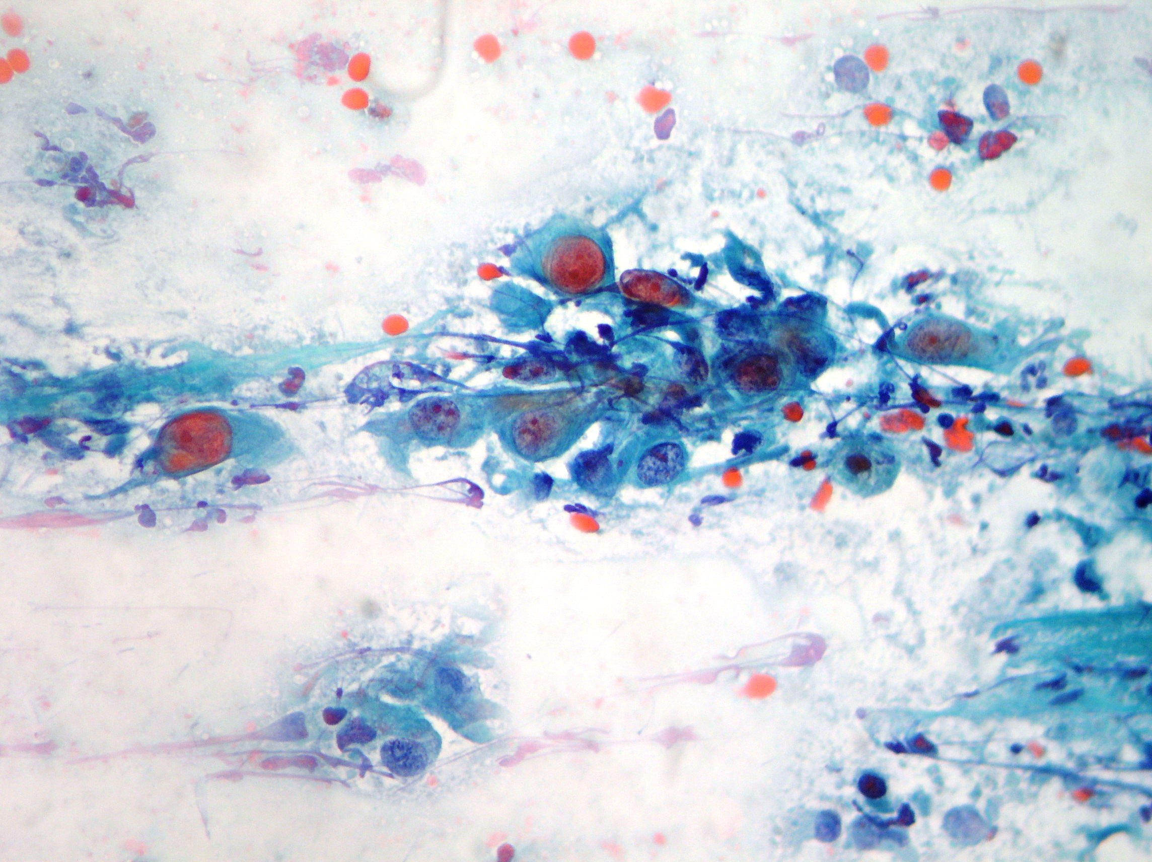
Lung adenocarcinoma cells
Cluster of tumor cells arranged in acini and papillary structures with macronucleoli and eccentric nuclei suggestive of lung adenocarcinoma. (Papanicolaou, x200)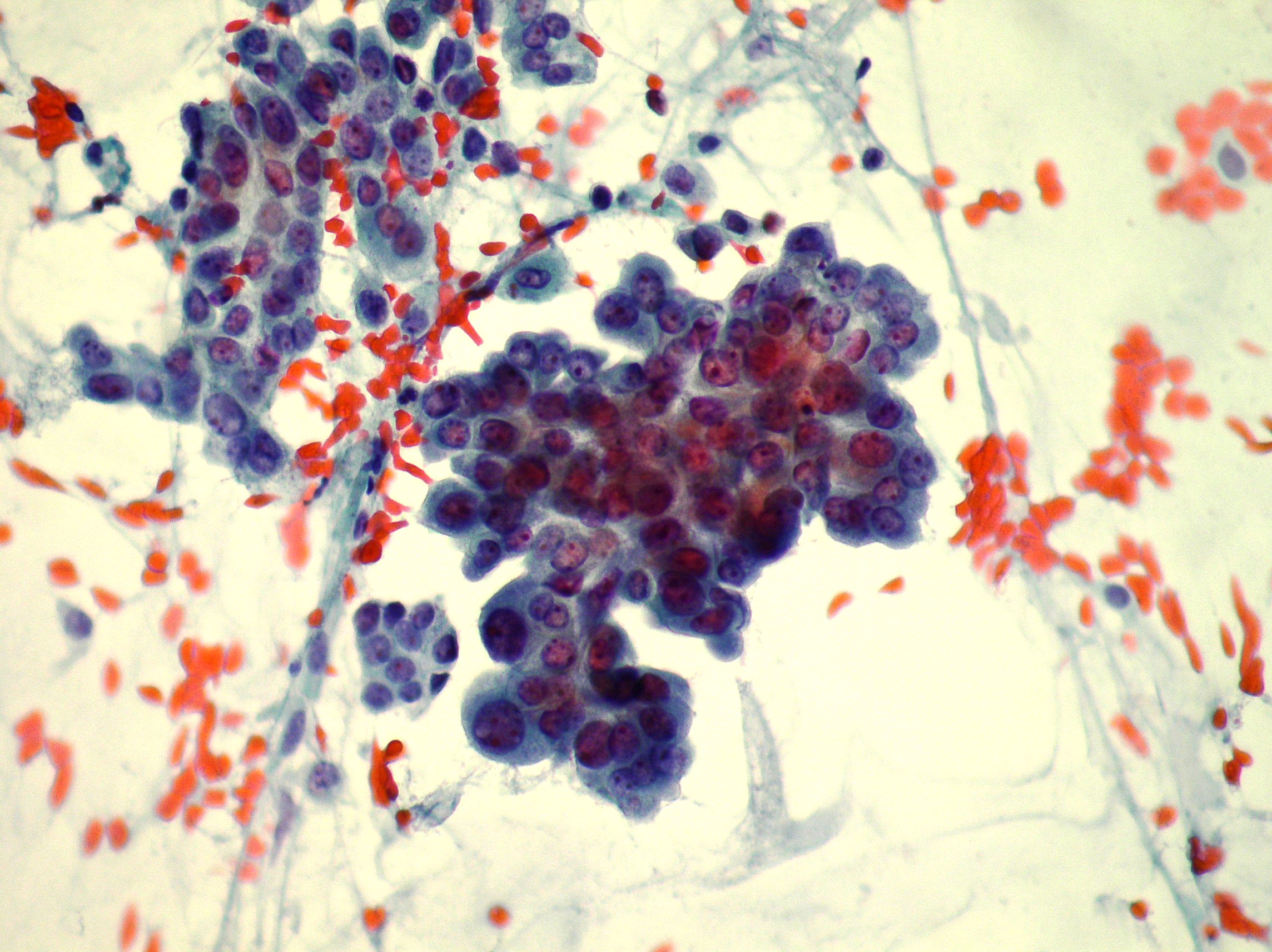
Normal ciliated bronchial cells
Normal ciliated bronchial cells obtained from bronchial brushing. (Papanicolaou, x200) 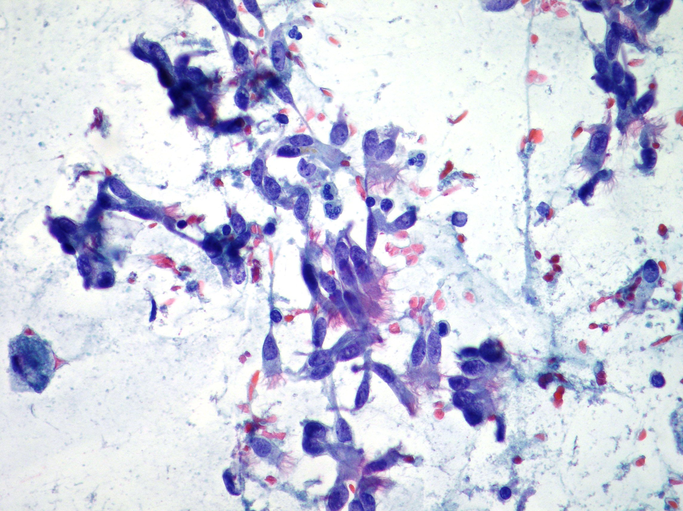
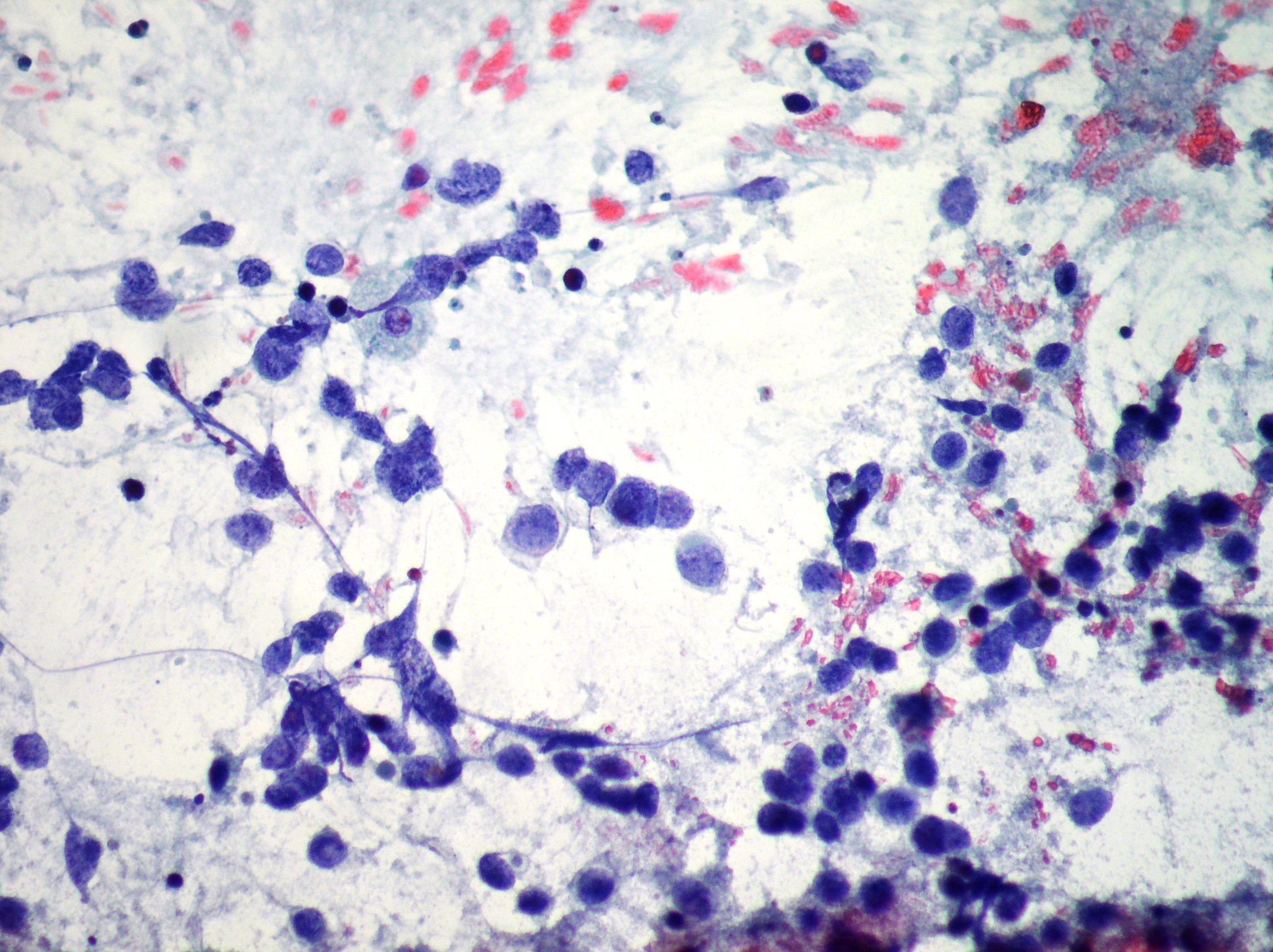
Small cell lung carcinoma
Oat cell type with nuclear molding, granular chromatin and high nucleocytoplasmic ratios. Necrosis in the background. Cytomorphology compatible with small cell lung carcinoma. (Papanicolaou, x200)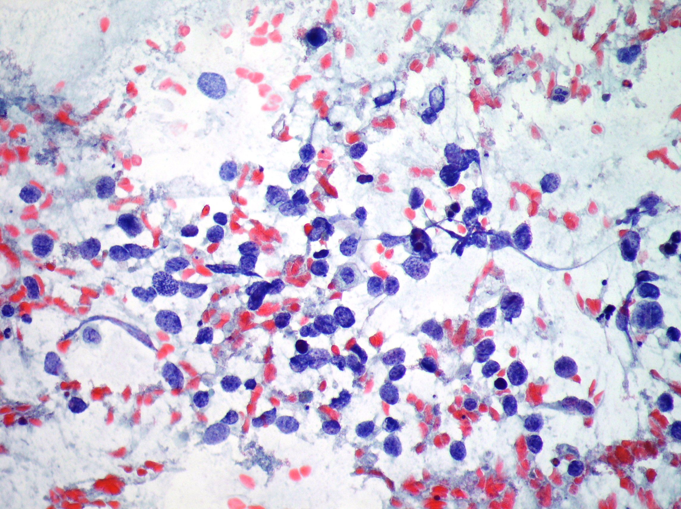
Keratinising squamous cell lung carcinoma
Keratinising squamous cell lung carcinoma with single cell presentation showing bizarre cell shape and irregular dense hyperchromatic nuclei. (Papanicolaou, x200)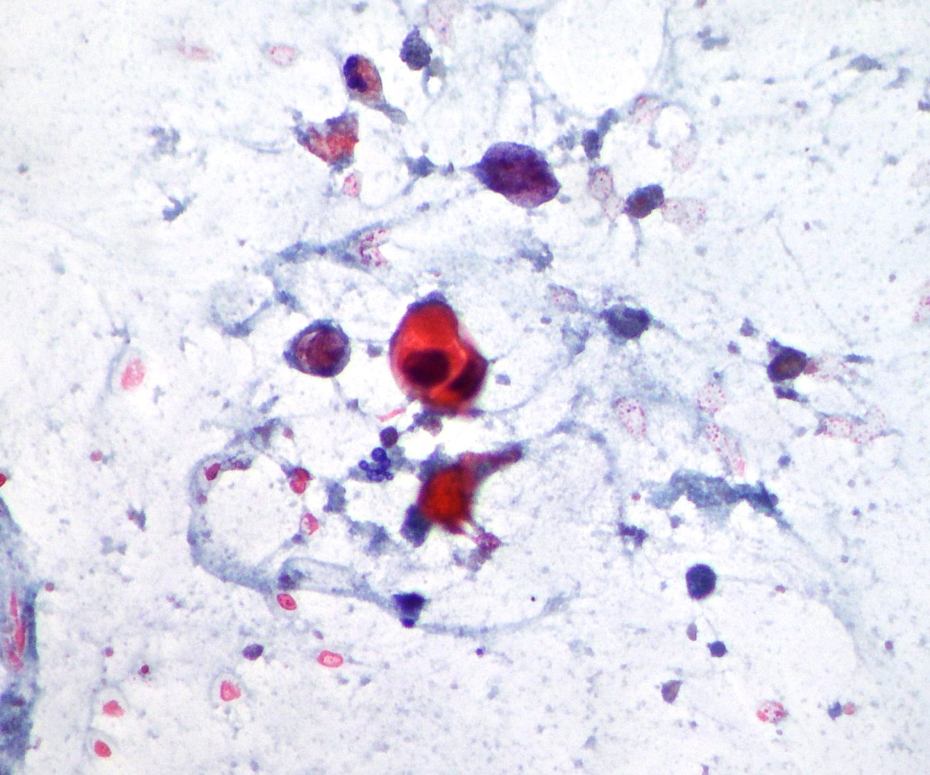
Non-keratinising squamous carcinoma.
Non-keratinising irregular cohesive fragment with irregular nuclear shape and chromatin distribution compatible with squamous cell lung carcinoma. (Papanicolaou, x200)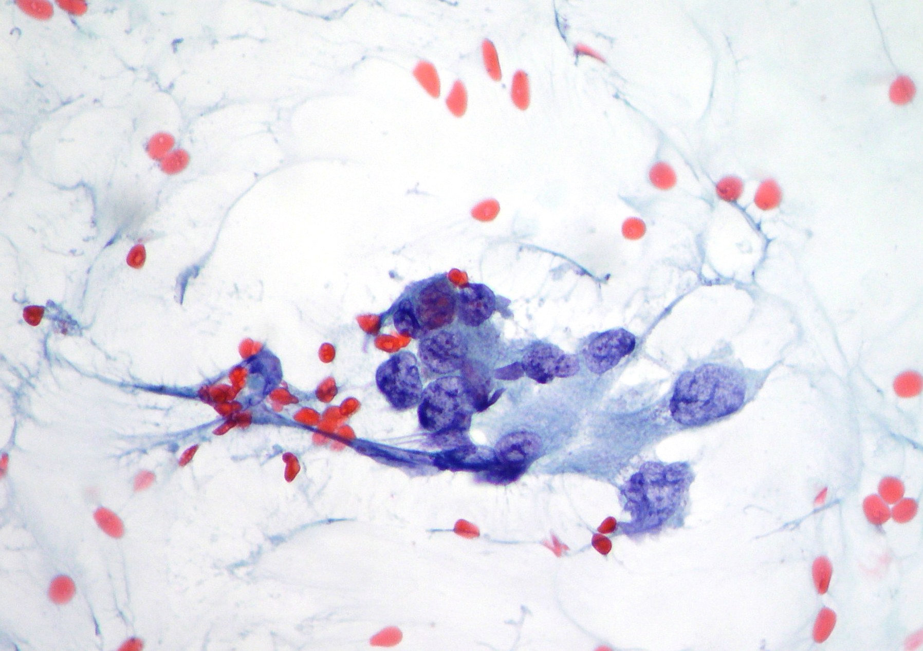
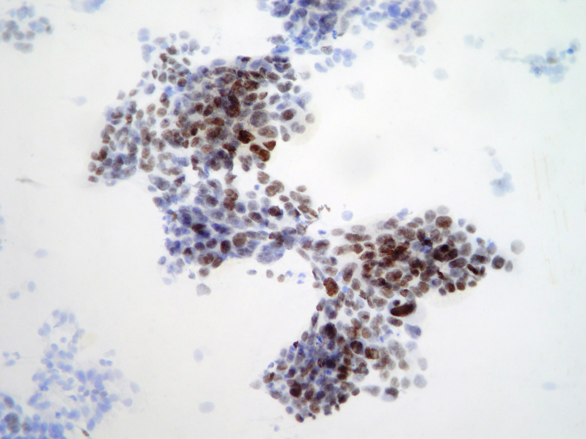
Lung adenocarcinama cells.
Sheets of neoplastic cells showing positive nuclear staining for TTF-1 compatible with lung adenocarcinama. (Papanicolaou, x200)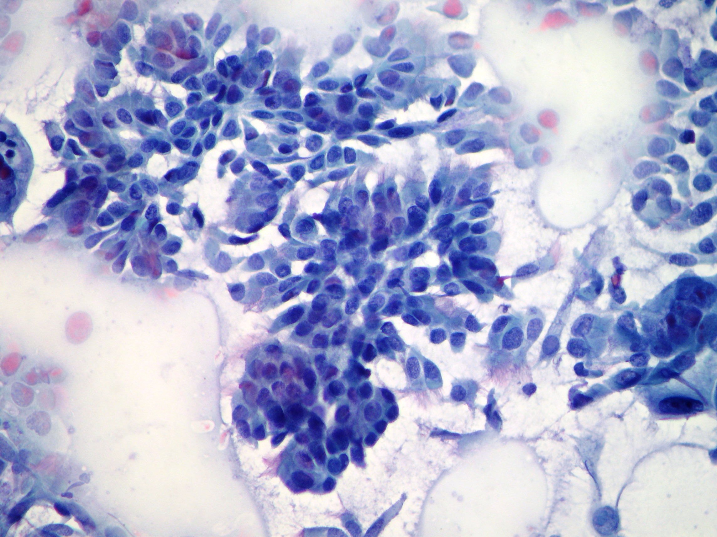
Non small cell lung cancer
Neoplastic single and in aggregate pleomorphic cells showing chromatin irregularly dispersed and hyperchromatic suggestive of non small cell lung cancer. (Papanicolaou, x200)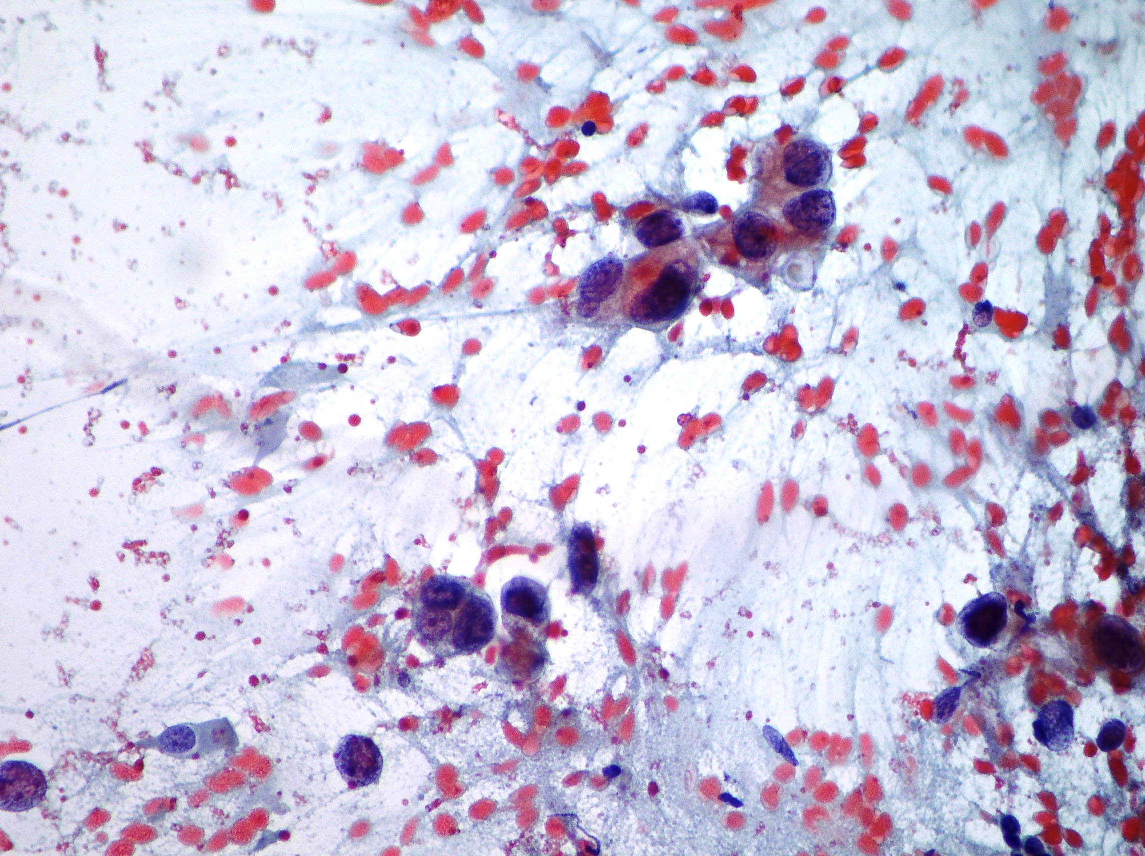
Lung adenocarcinoma
Papillary structure showing variability in amount of pleomorphism and nuclear atypia, micro-macrocalcifications and cell debris on the background resulted suggestive of lung adenocarcinoma. (Papanicolaou, x200)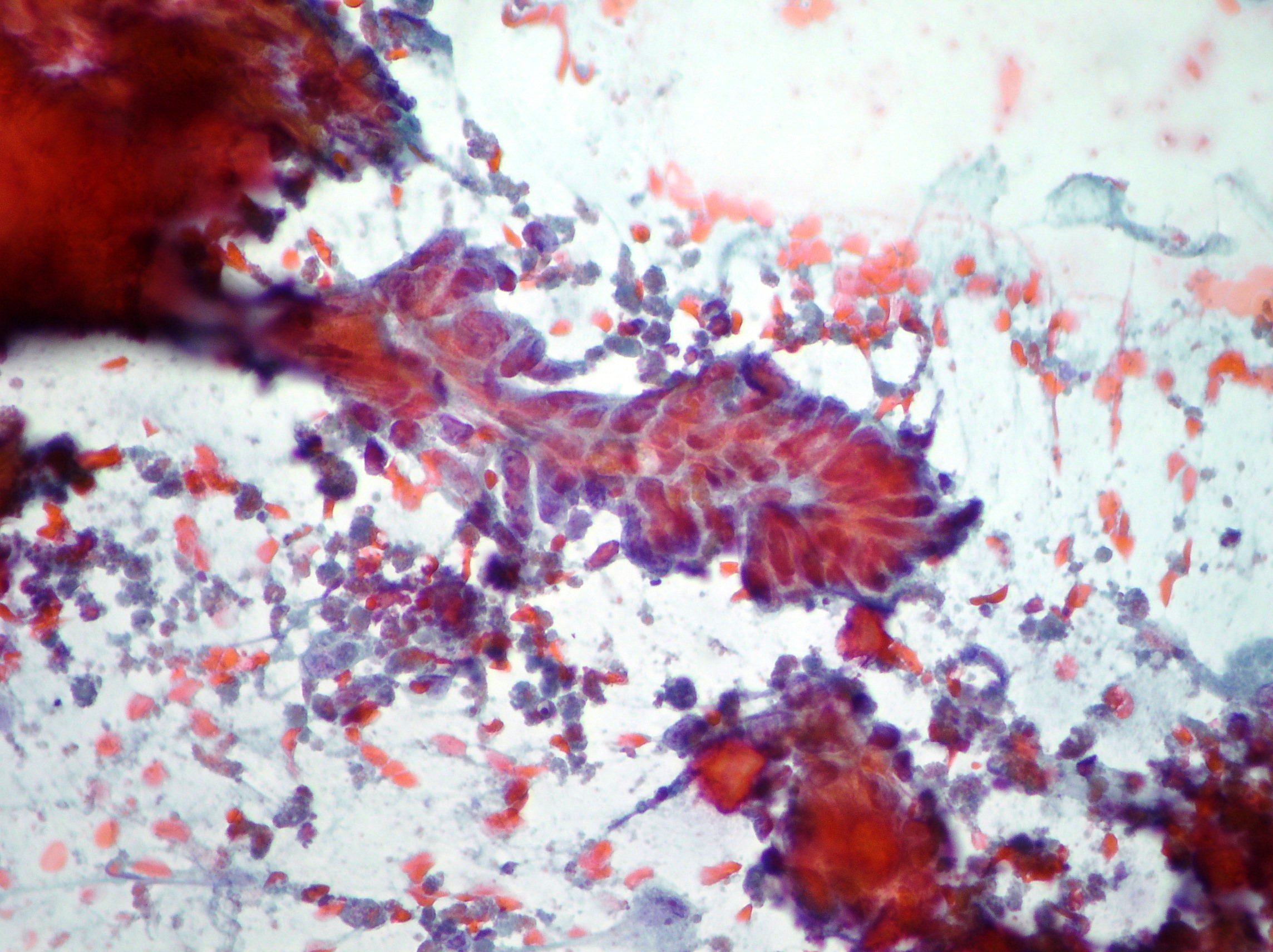
Papillary adenocarcinoma
Papillary cell cluster suggestive of adenocarcinoma differentiation. (Papanicolaou, x400)