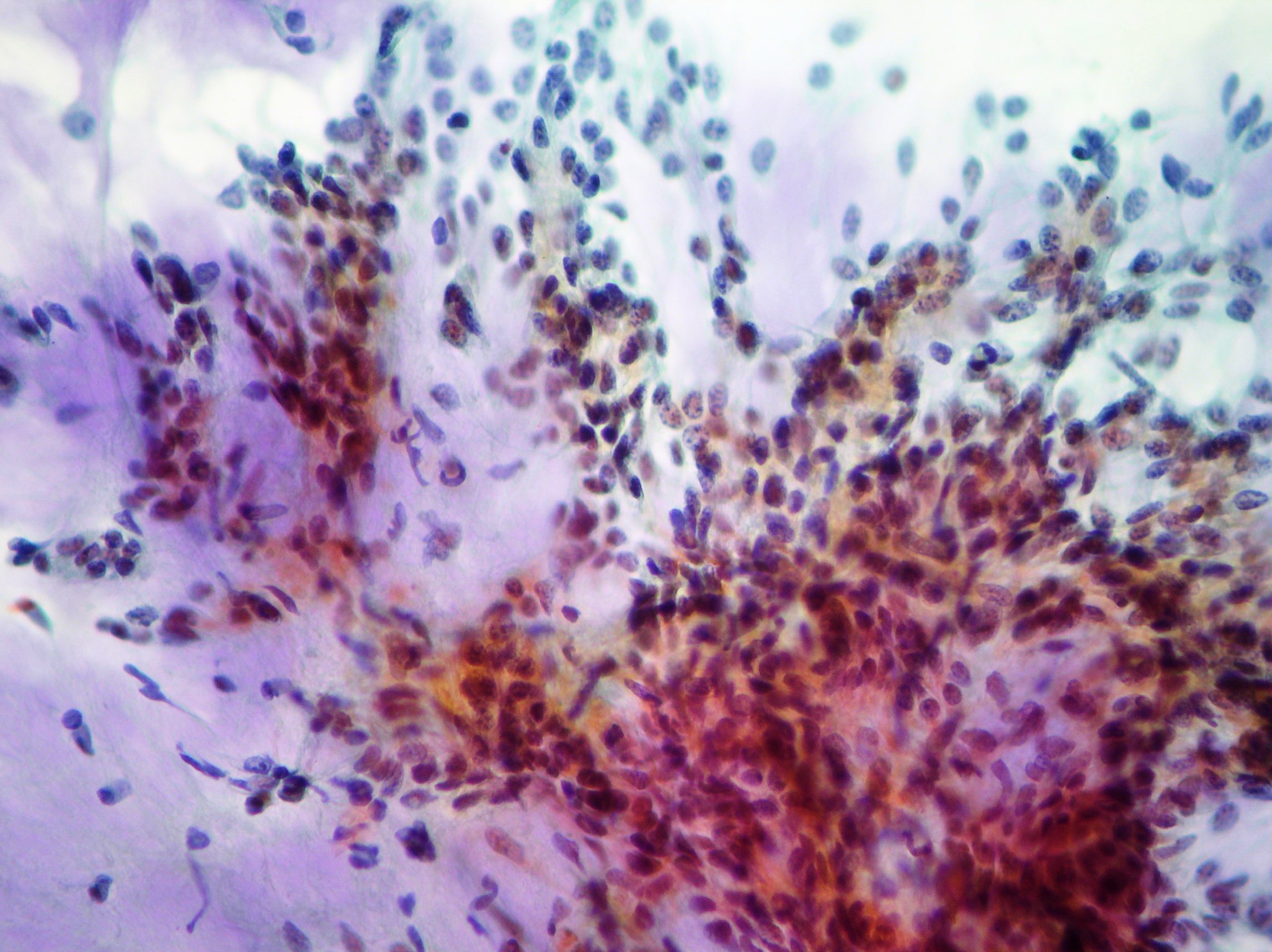
Acinar cells neoplasia
FNAC. Large cells with small basal nuclei arranged in alveolar aggregates and cytoplasm clear and vacuolized suggestive of acinar carcinoma. Histopathological verification is necessary (DiffQuick and Papanicolaou staining x100, x200, x100)
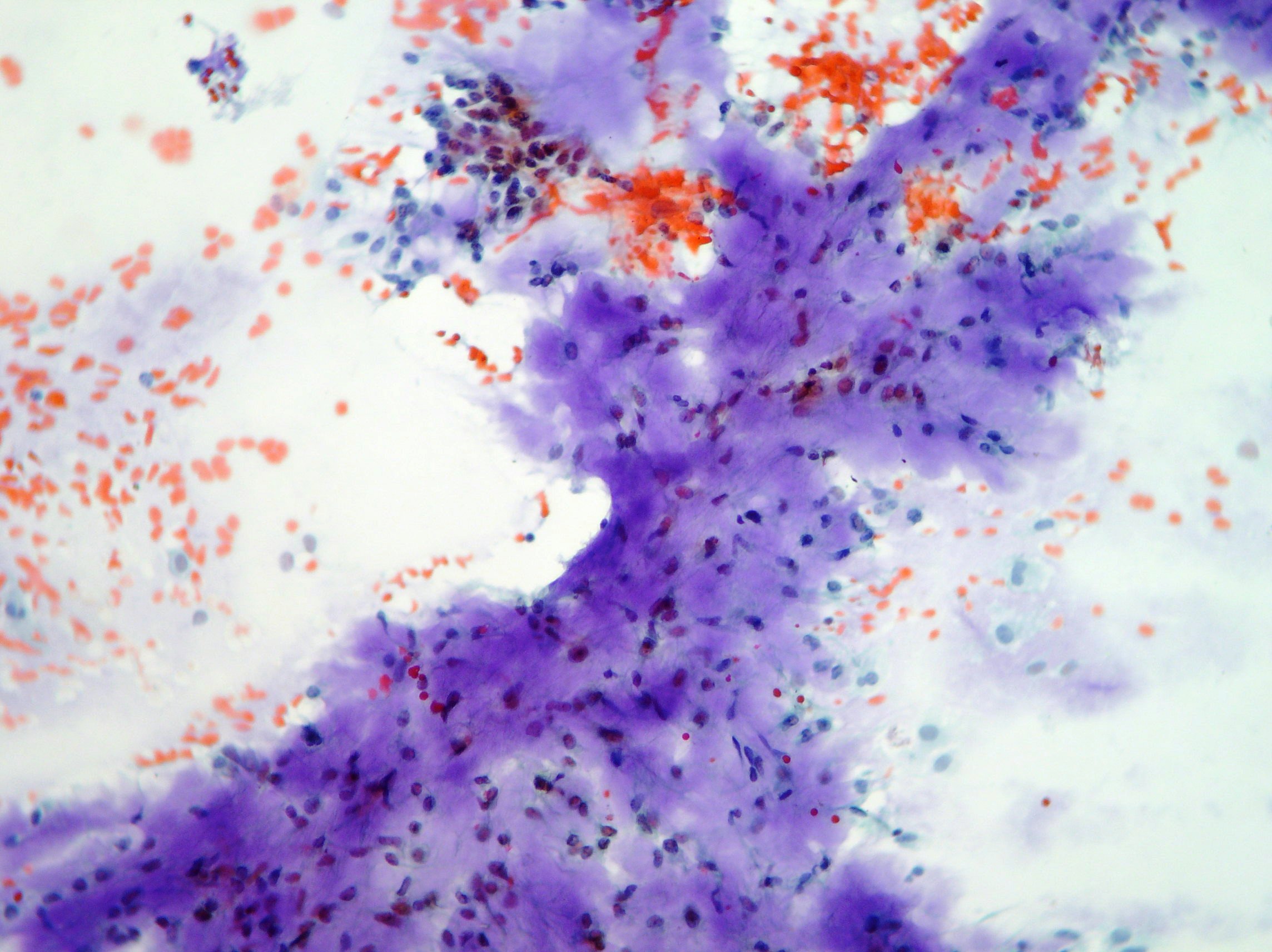
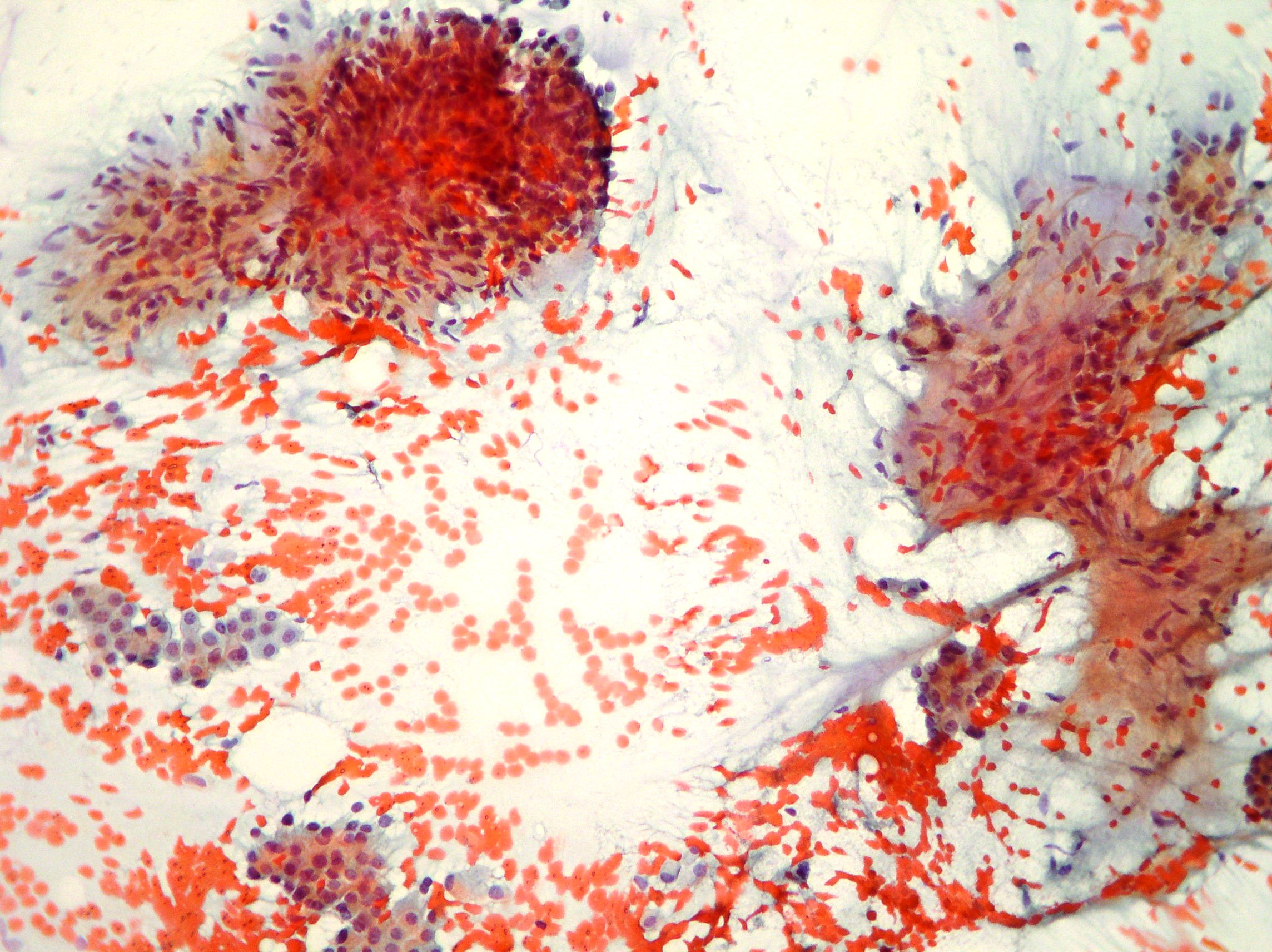
Pleomorphic adenoma
FNAC. Pleomorphic adenoma showing epithelial-like cells associated to fibrillar fibromyxoid stroma including oval and spindle cells. (Papanicolaou, x200, x100, x100)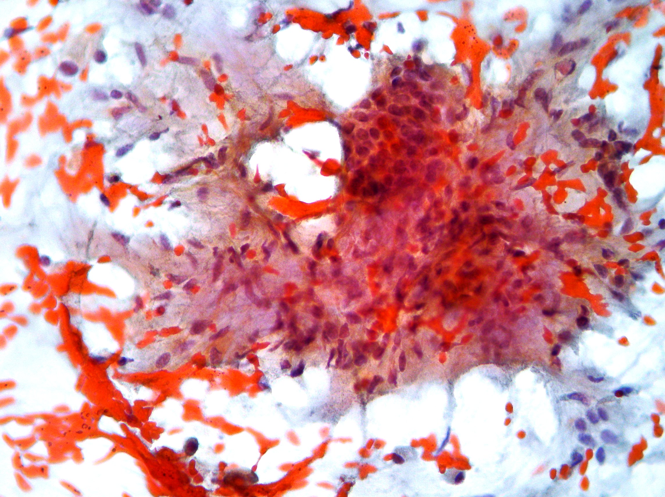
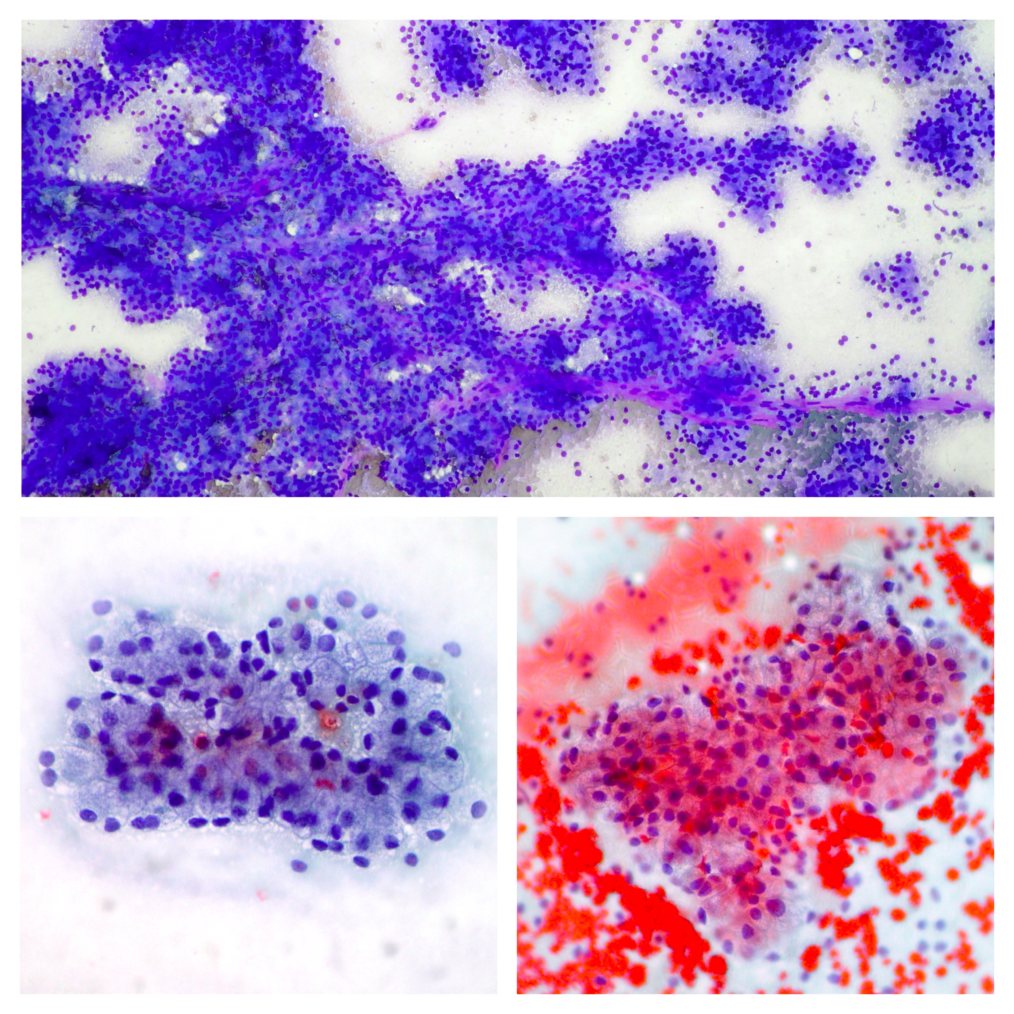
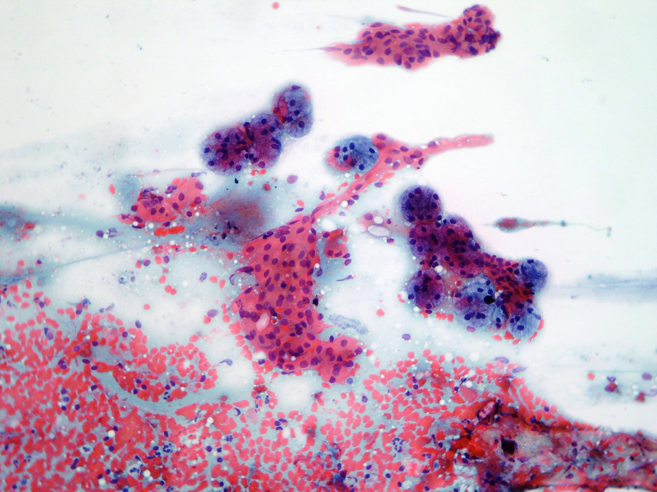
Warthin’s tumor
Warthin’s tumor showing groups of uniform oncocytic epithelial cells with small nuclei, lymphocytes, normal acini and proteinaceous material in the background. (Papanicolaou, x200, x100)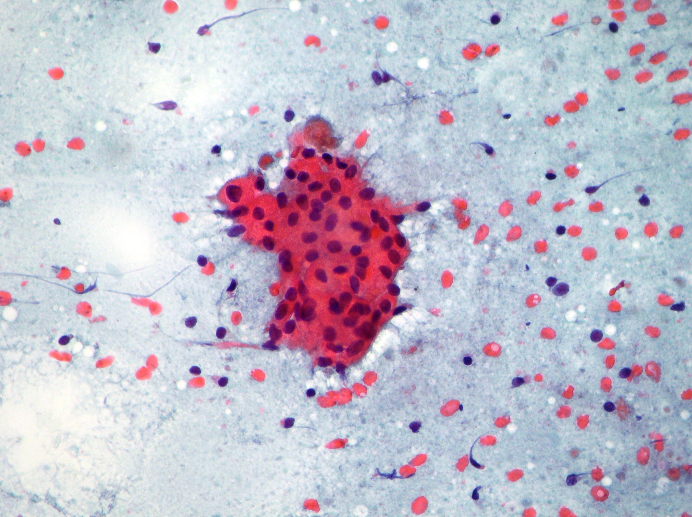
Oncocytic neoplasia
Aggregates of cohesive oncocytic cells with small and regular nuclei suggestive of oncocytic neoplasia. (Papanicolaou, x100)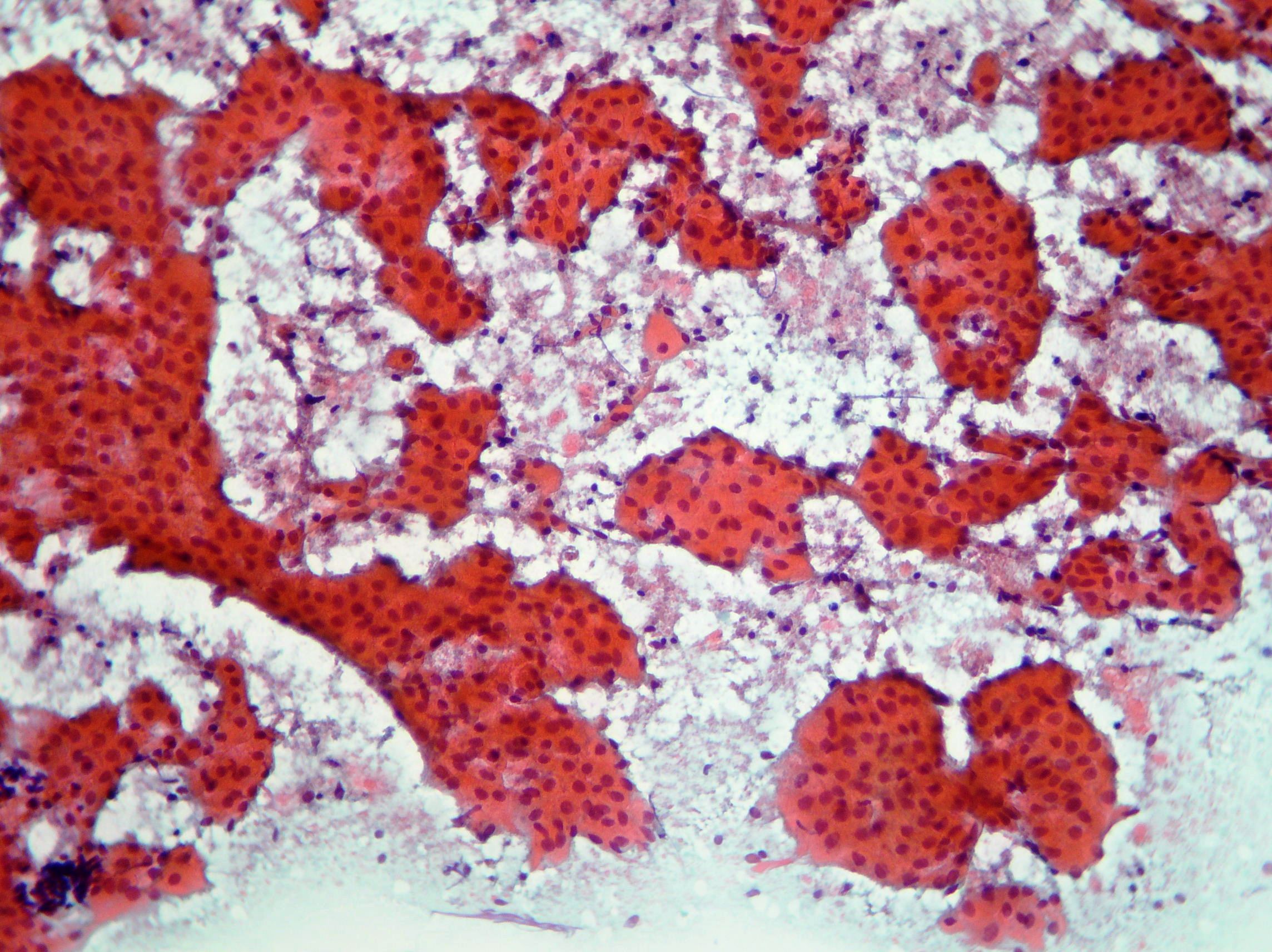
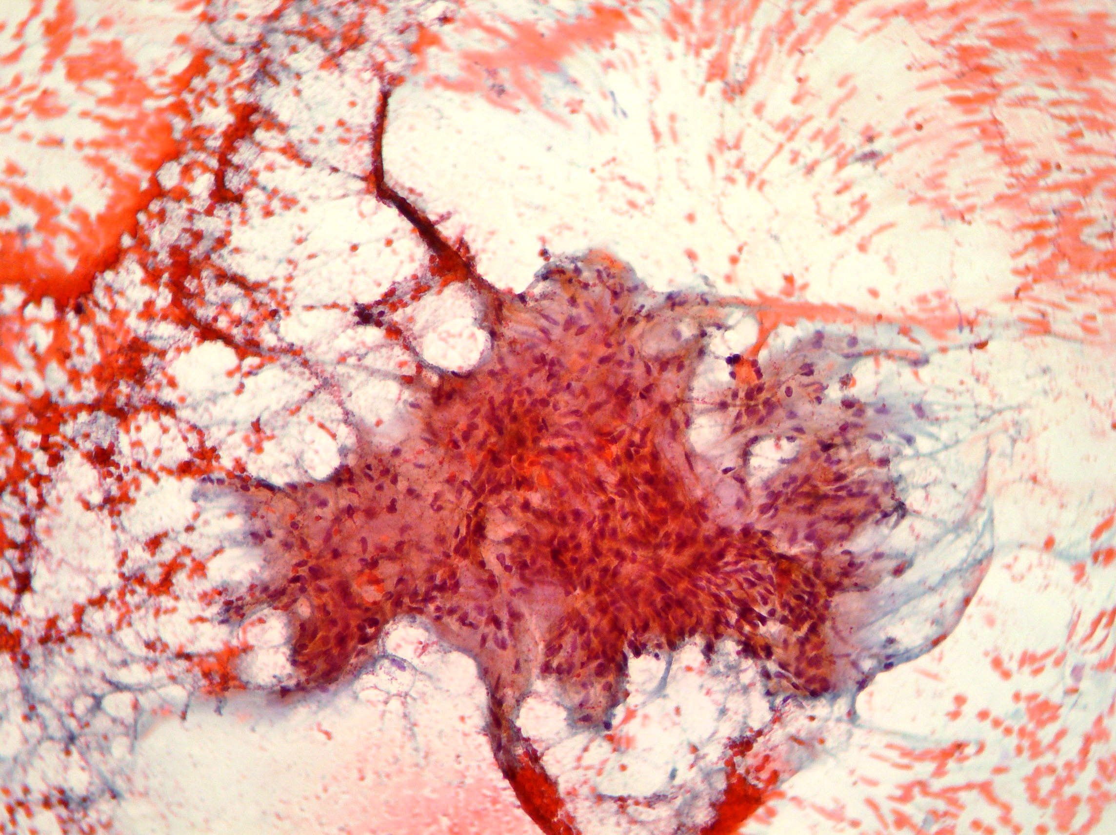
Pleomorphic adenoma
Pleomorphic adenoma showing typical fibrillary chondromyxoid substance. (Papanicolaou x200, x100)