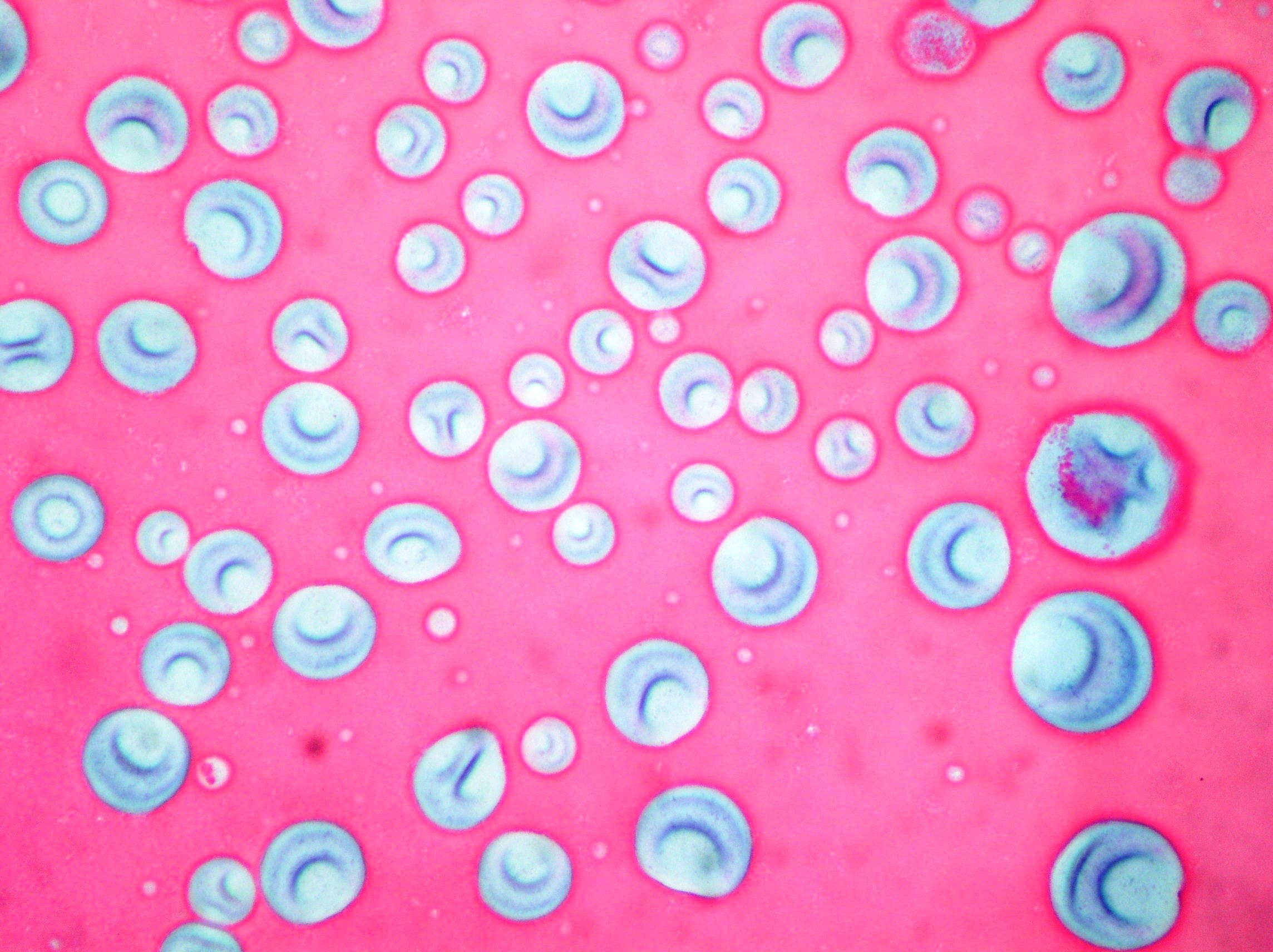
Colloid
Air bubbles and colloid background from a thyroid goiter. (Papanicolaou, x100)
Anaplastic carcinoma
Bizzarre malignant cells with mitotic figures, macronucleoli, debris and inflammatory background(Papanicolaou x200)

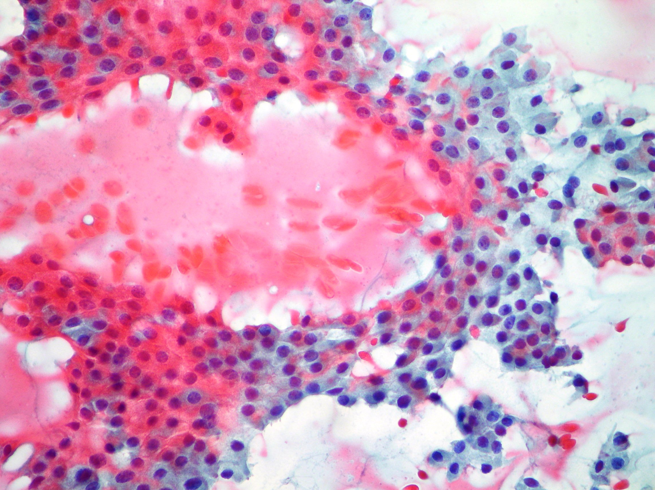
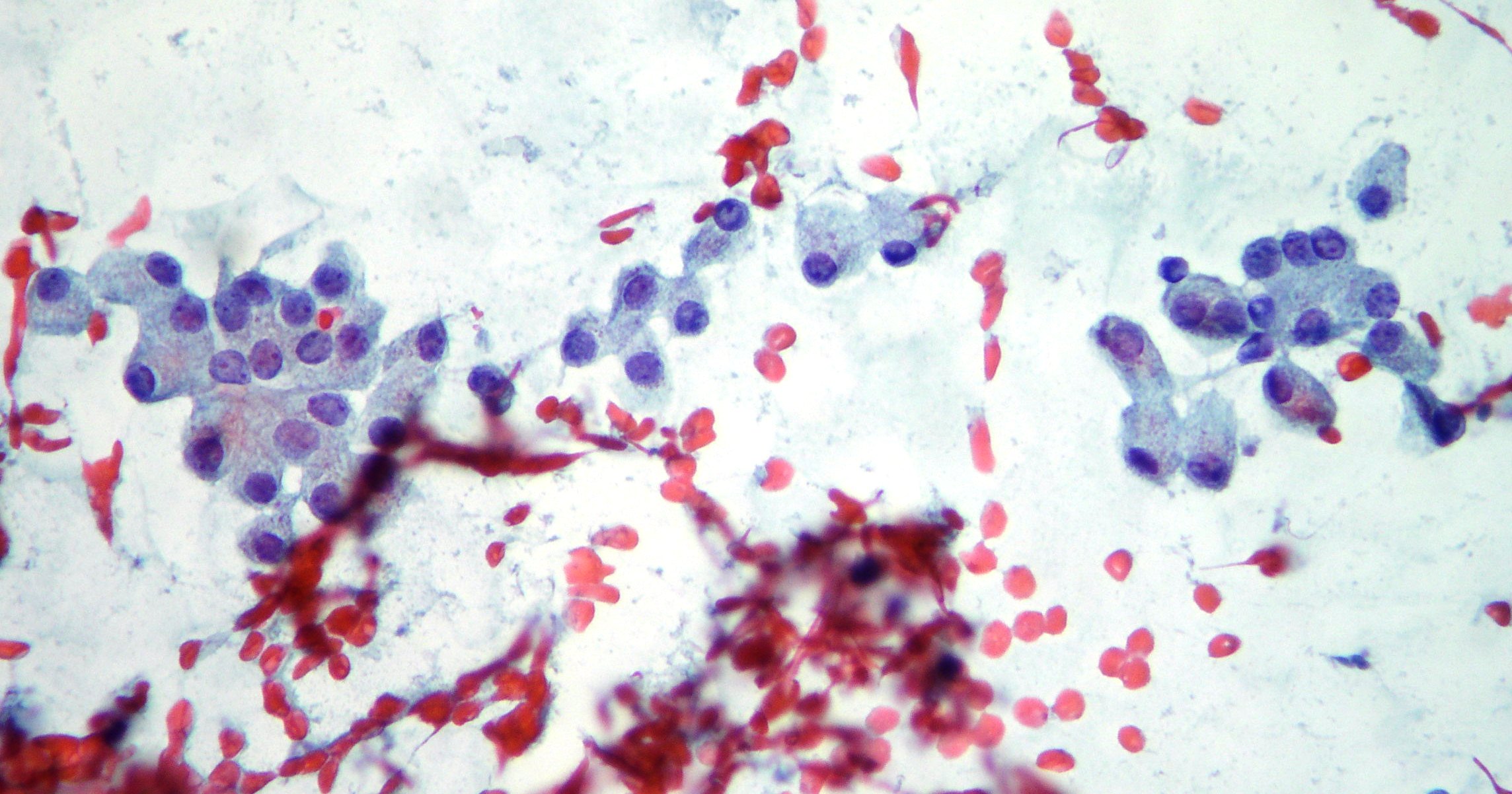
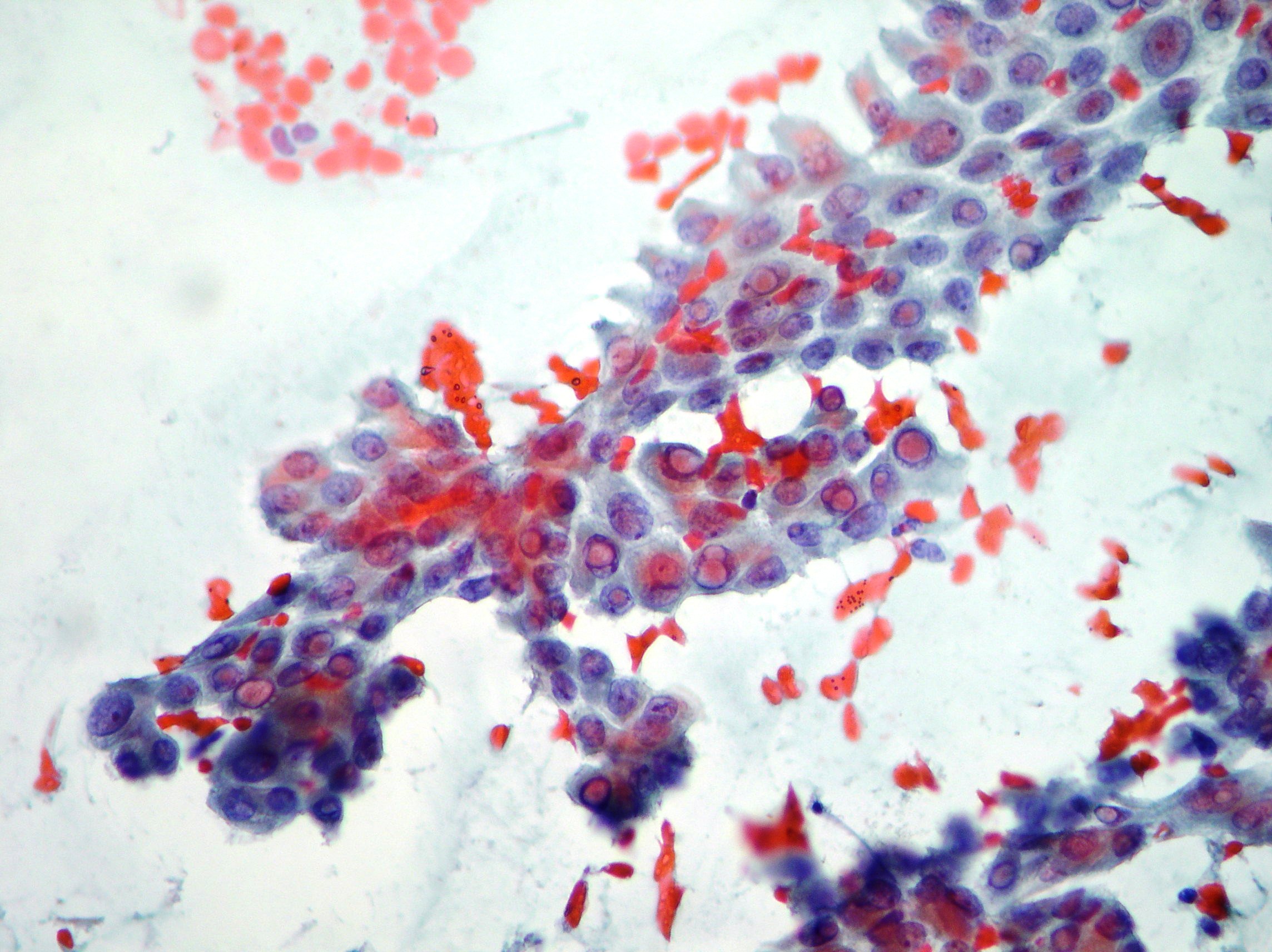
Papillary carcinoma
Fragments and groups of thyroid papillary carcinoma showing disorderly and overlapping arrangement of oxyphil cells sometimes multinucleated with pseudo inclusions. (Papanicolaou, x100, x200)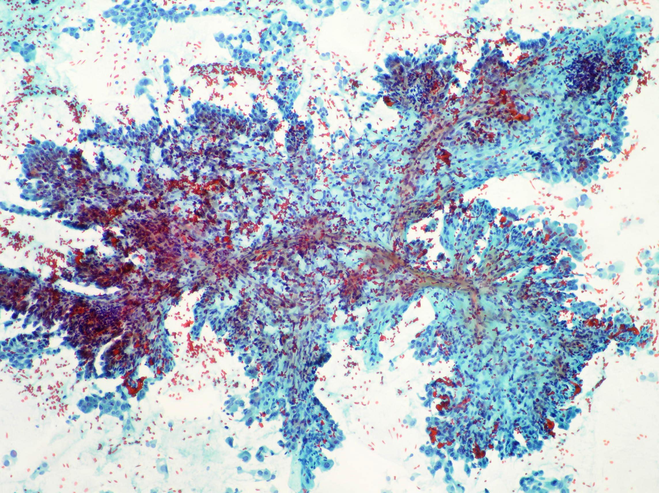
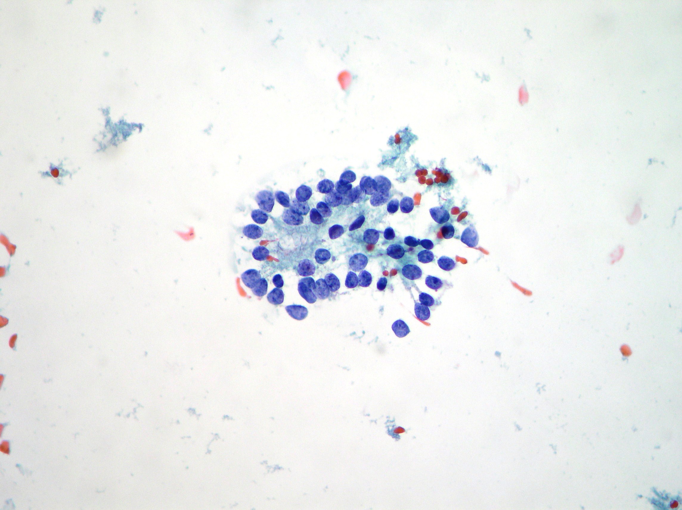
Follicular proliferation and thyroiditis
A case of thyroiditis associated to follicular proliferation showing micro follicular cell clusters and rosettes in absence of colloid. (Papanicolaou x100, x200) 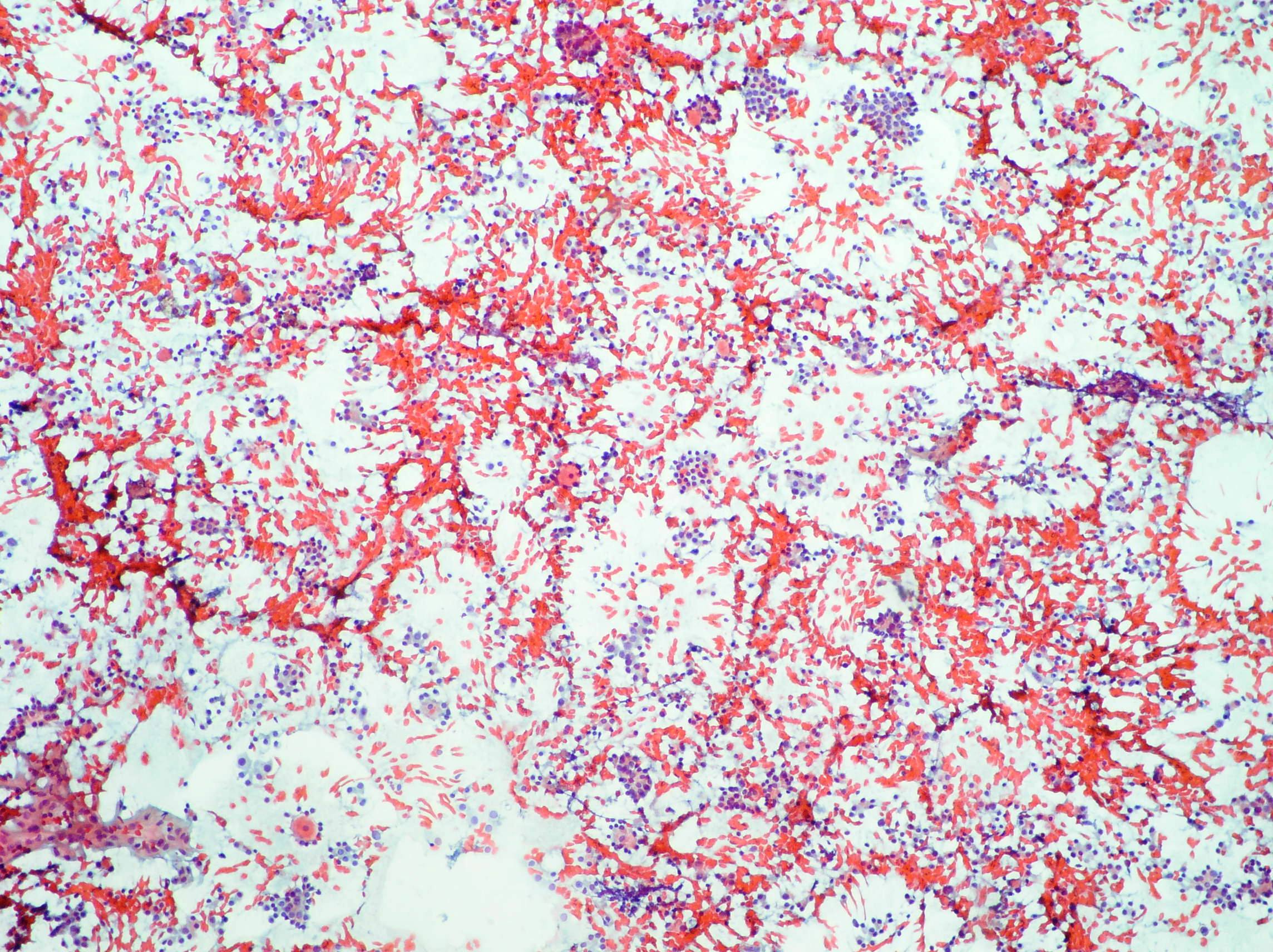
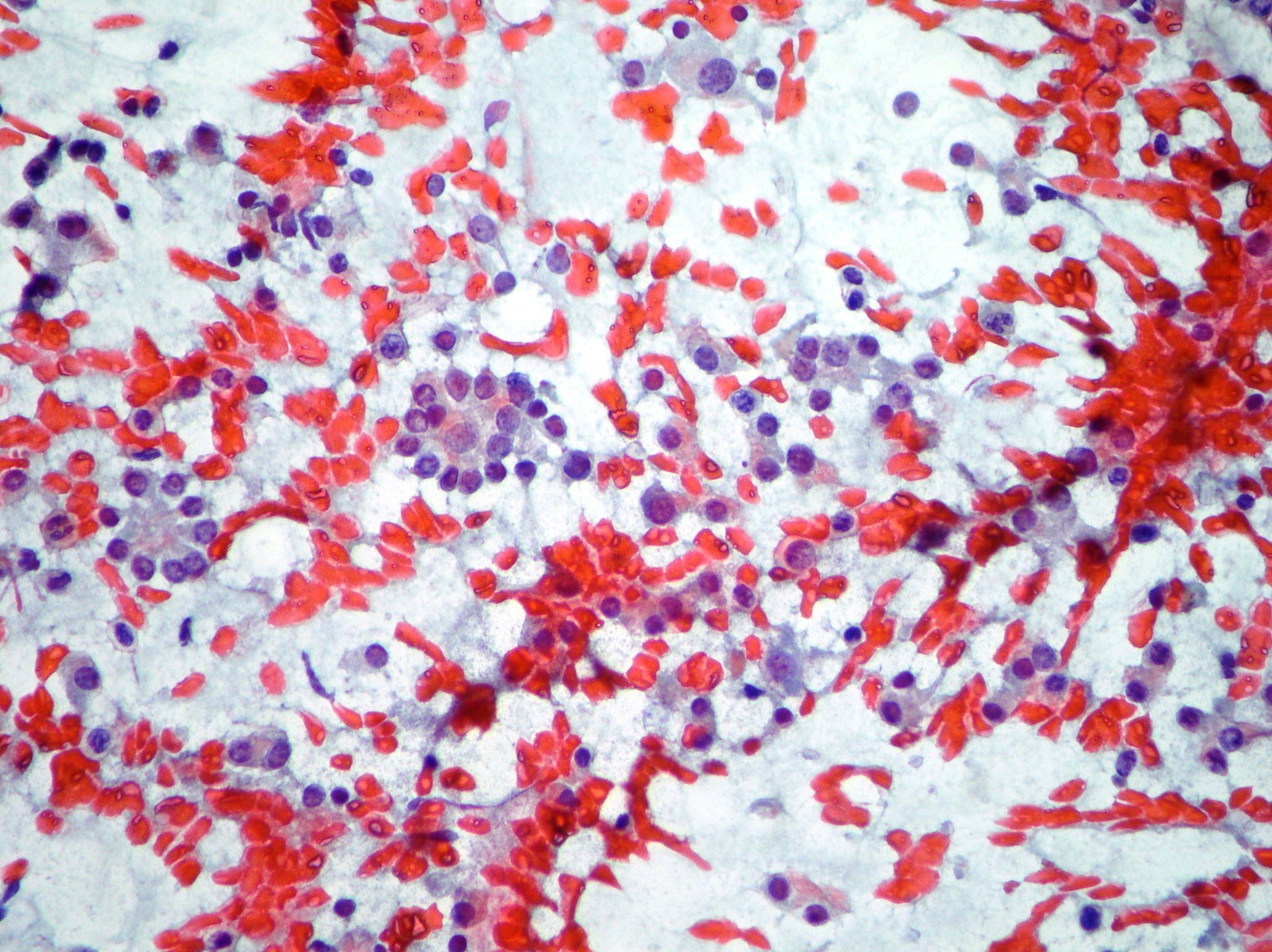
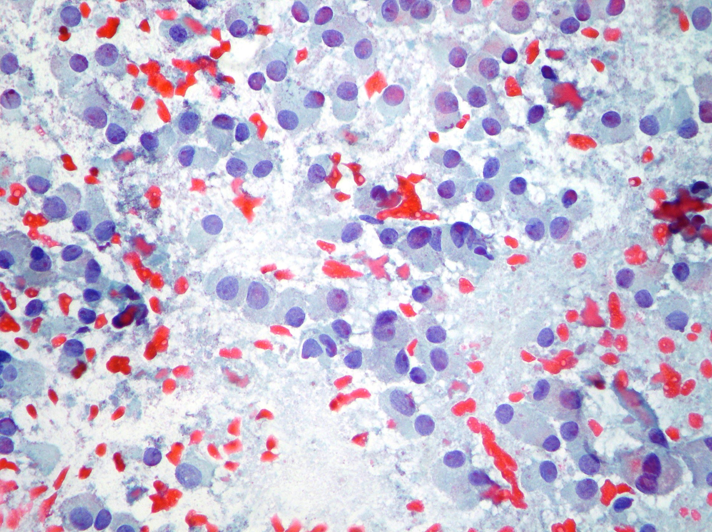
Medullary carcinoma
Thyroid medullary carcinoma showing dispersed and clustering cells with “plasmacytoid” appearance, moderate anisokariosis and binucleations. (Papanicolaou x 200) 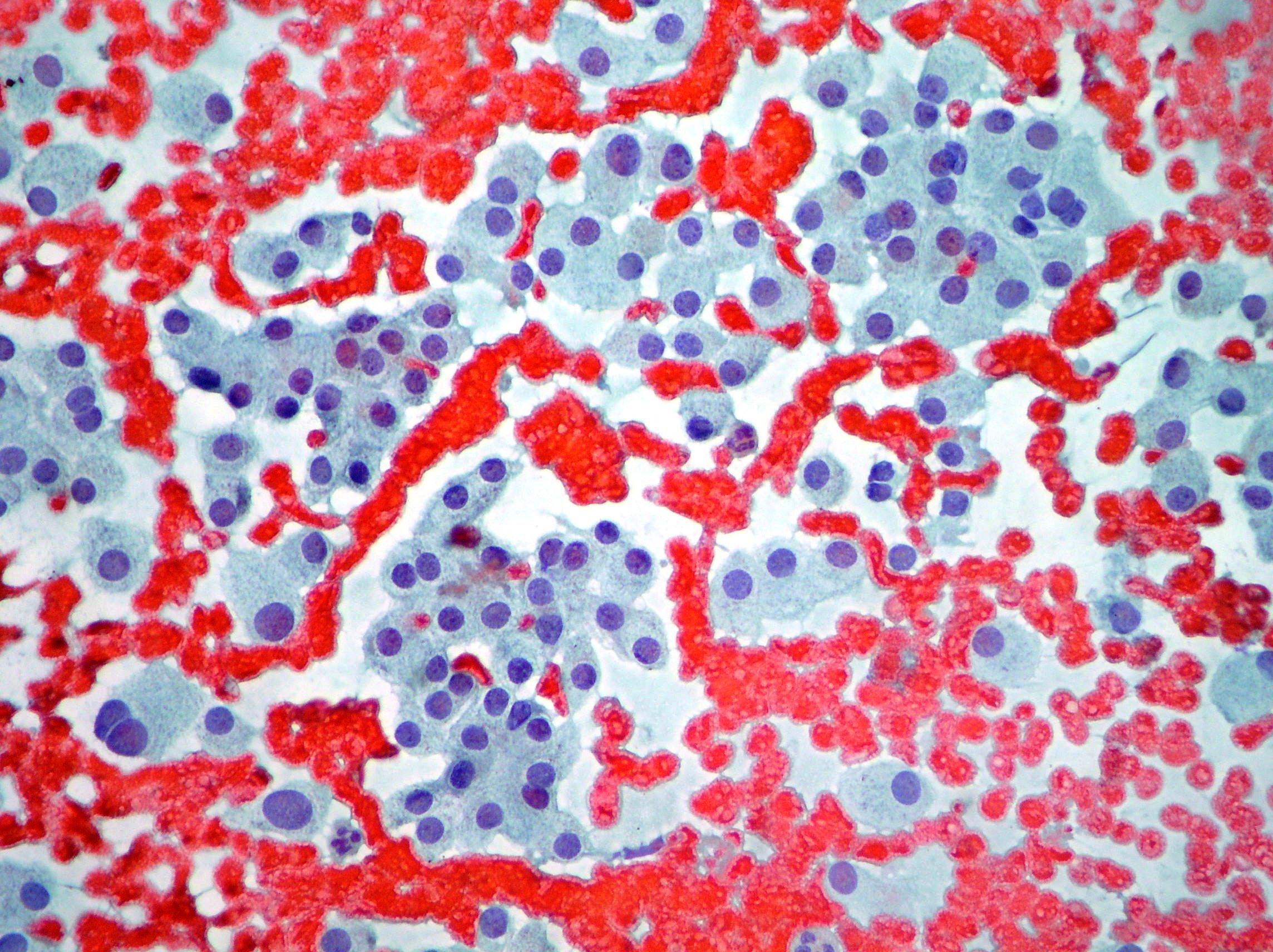
Oxyphil (Hurtle cell) proliferation
Oxyphil cells proliferation: histology oxyphil adenoma (Papanicolaou x100, x200) 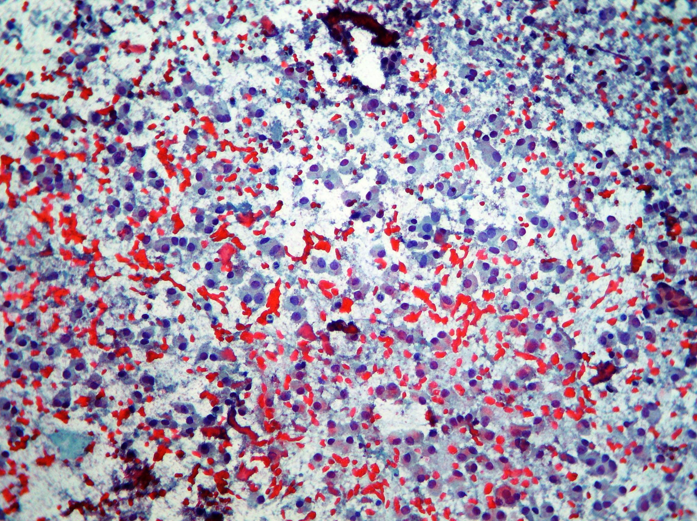
Colloid
Cracking artefacts of colloid. (Papanicolaou, x100)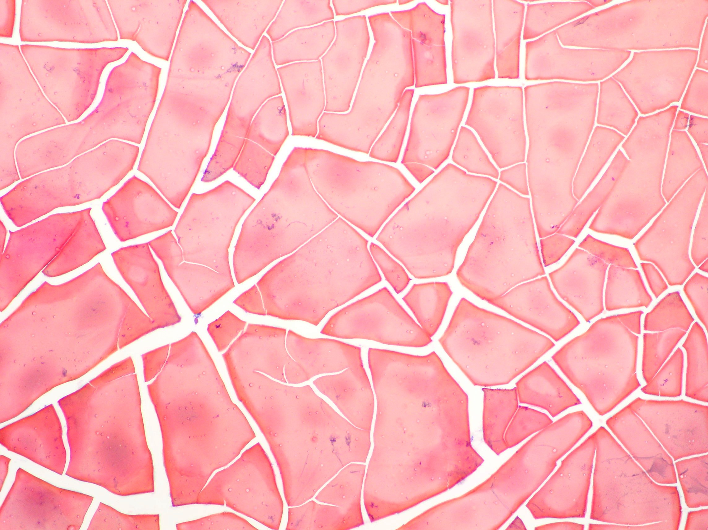
Oxyphil proliferation
Oxyphil cell proliferation showing clusters of relatively small cells in abundant and widespread colloid. (Papanicolaou, x100, x400) 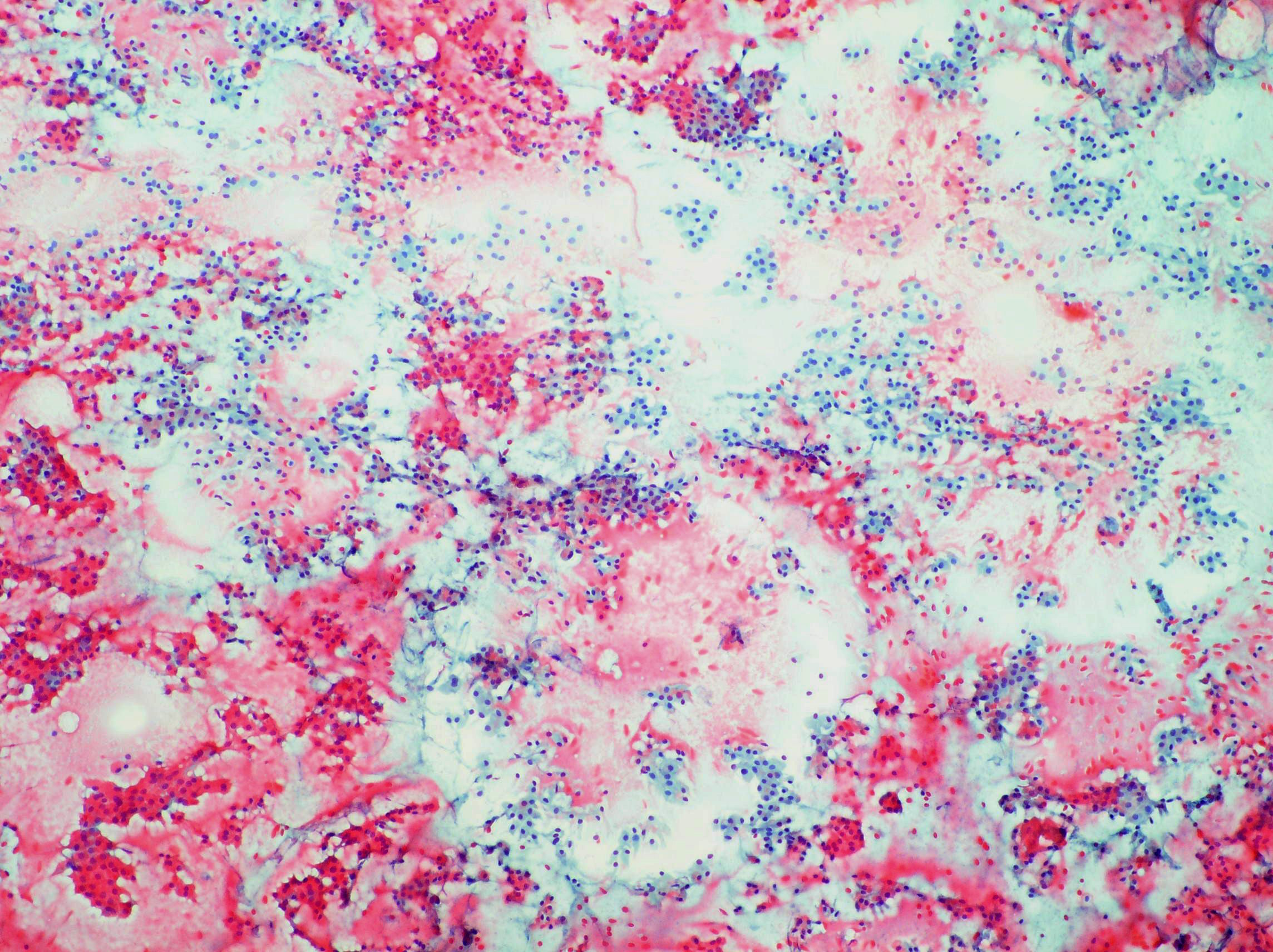
Papillary carcinoma
Monolayered sheet of papillary carcinoma showing disorderly and overlapping arrangement of cells and nuclei with some pseudoinclusions. (Papanicolaou, x200)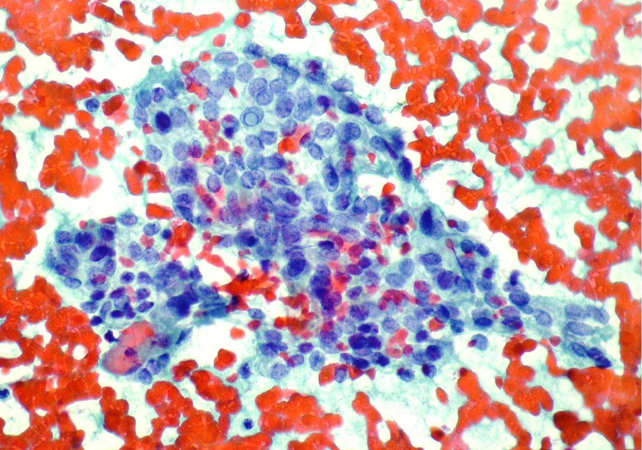
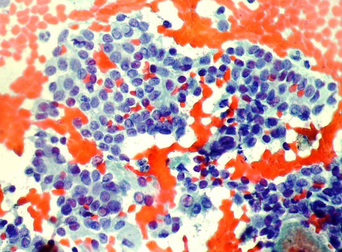
Medullary carcinoma
Cellular smear showing isolated and arranged monolayer epithelial cells. Round, triangular, plasmacytoid, mono- and binucleated cells with moderate cytoplasm suggestive of medullary carcinoma. (Papanicolaou, x200) 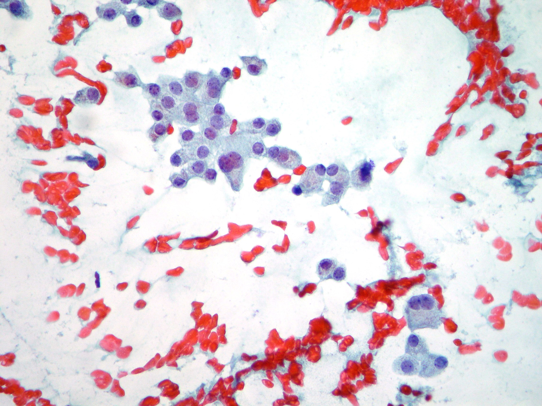
Hyperplasia
Flat monolayer sheet of epithelial cells with uniformly small round nuclei in a honeycomb structure associated with abundant colloid, compatible with colloid goiter and hyperplasia. (Papanicolaou, x100) 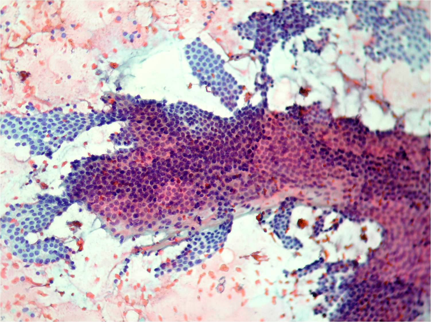
Normal thyreocytes
Normal epithelium showing uniform thyrocytes with partially disrupted cytoplasm in a colloid and ematic background. (Papanicolaou, x400) 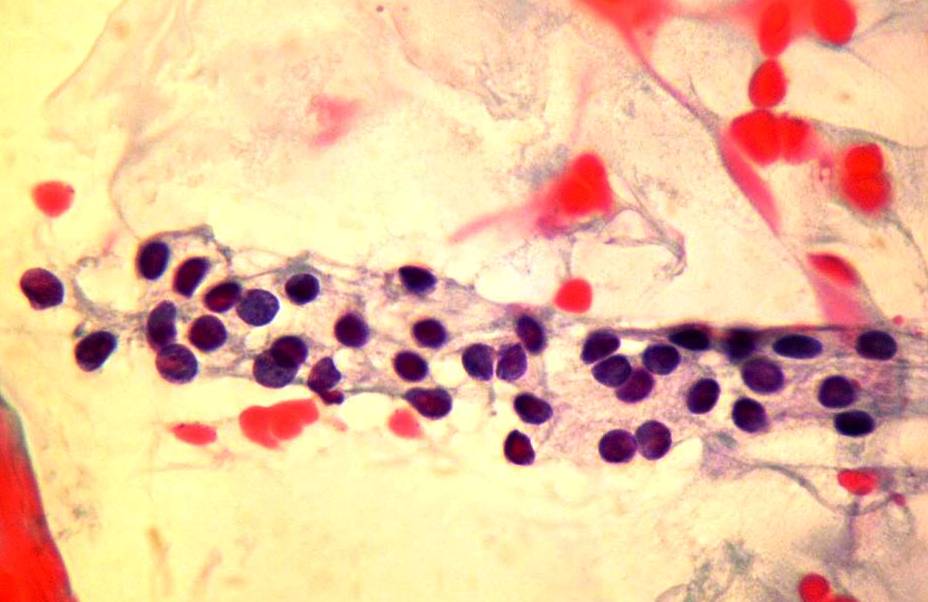
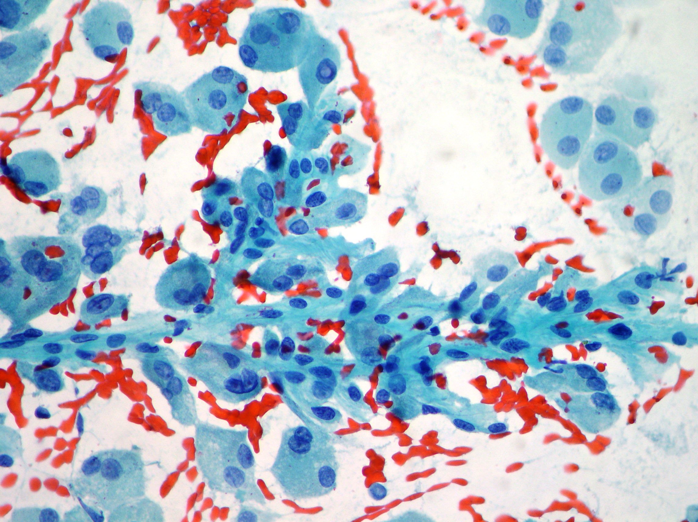
Papillary carcinoma.
Sheet of cells showing many intranuclear vacuoles. (Papanicolaou, x400)
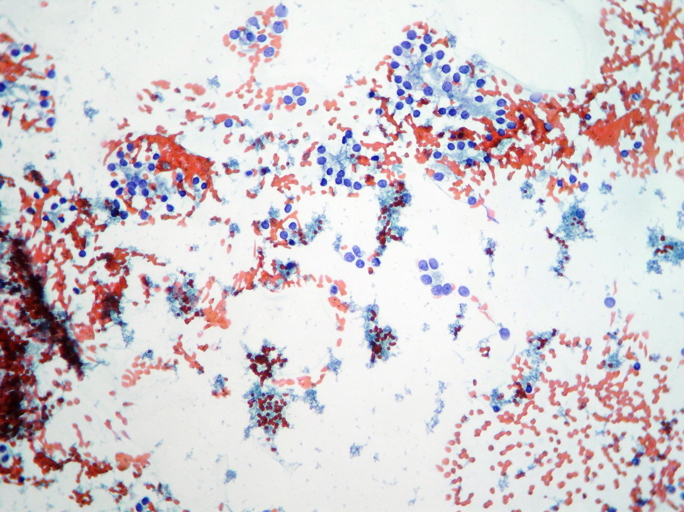
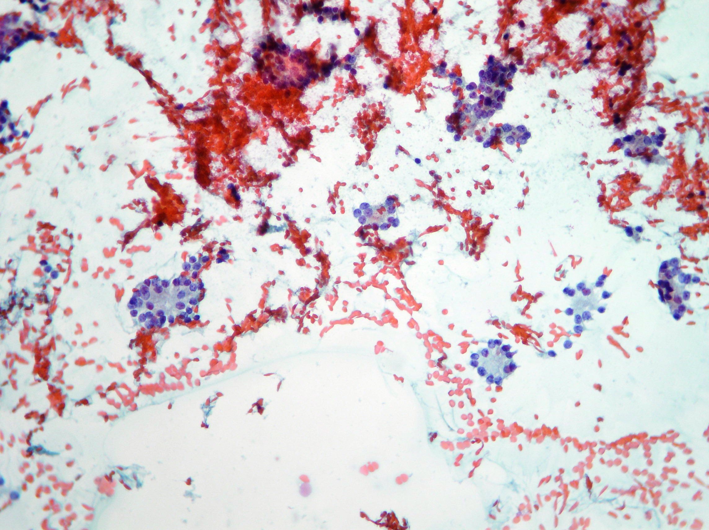
Thyroiditis
Florid lymphocytic thyroiditis showing cellular smears of mainly reactive mixed lymphoid cell population. (Papanicolaou, x200) 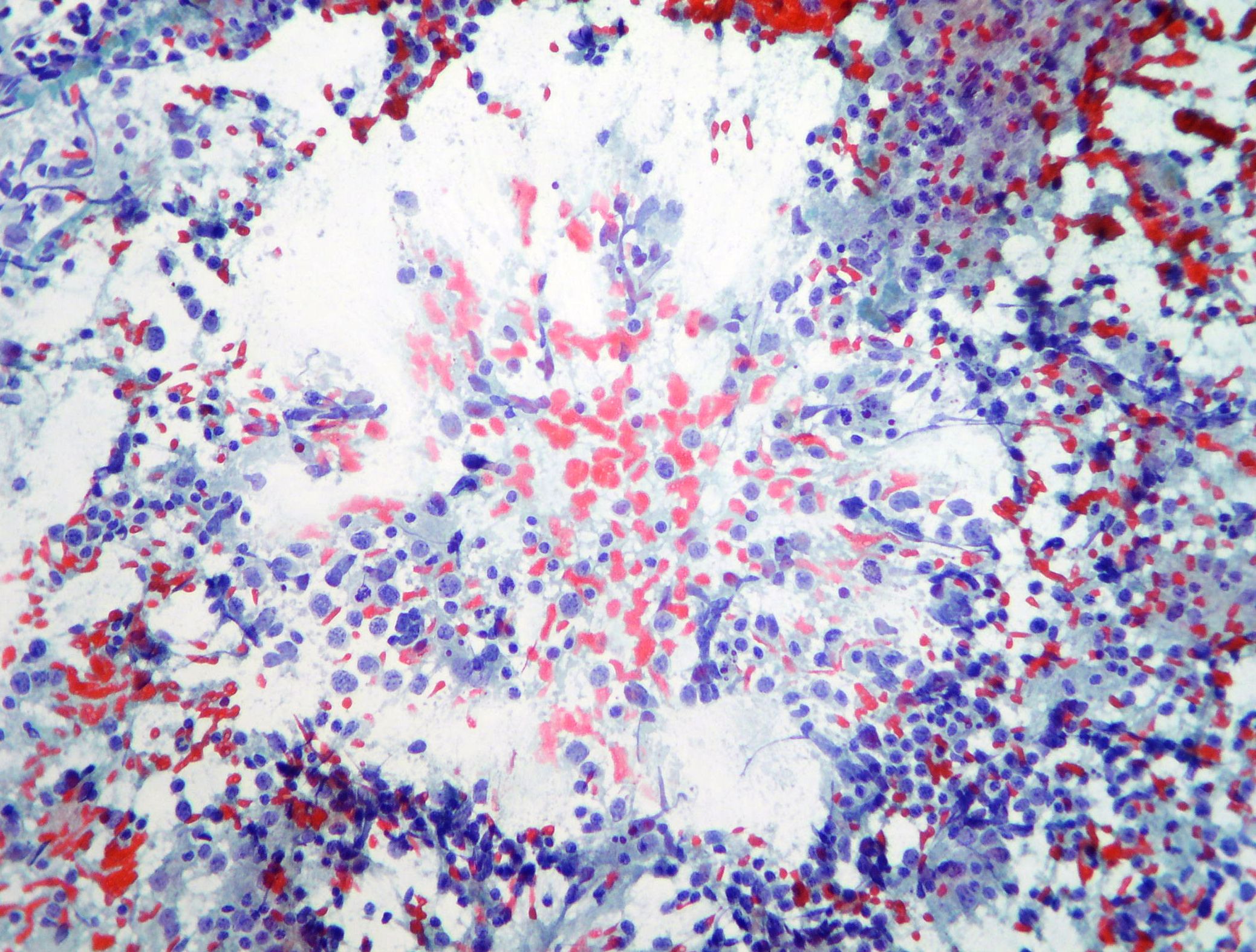
Papillary cell carcinoma
Fragment of micro-architecture showing papillae with a fibrovascular core compatible with papillary cell carcinoma. (Papanicolaou, x100)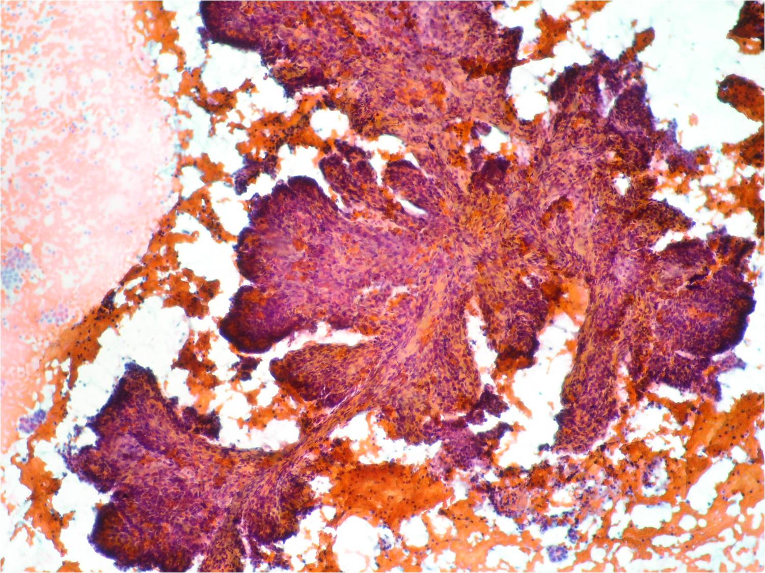
Hyperplasia
FNAC. Large flat monolayered sheet, groups and single thyroid cells showing monotonous sizes on abundant colloid suggestive of adenomatous hyperplasia. (Papanicolaou, x100)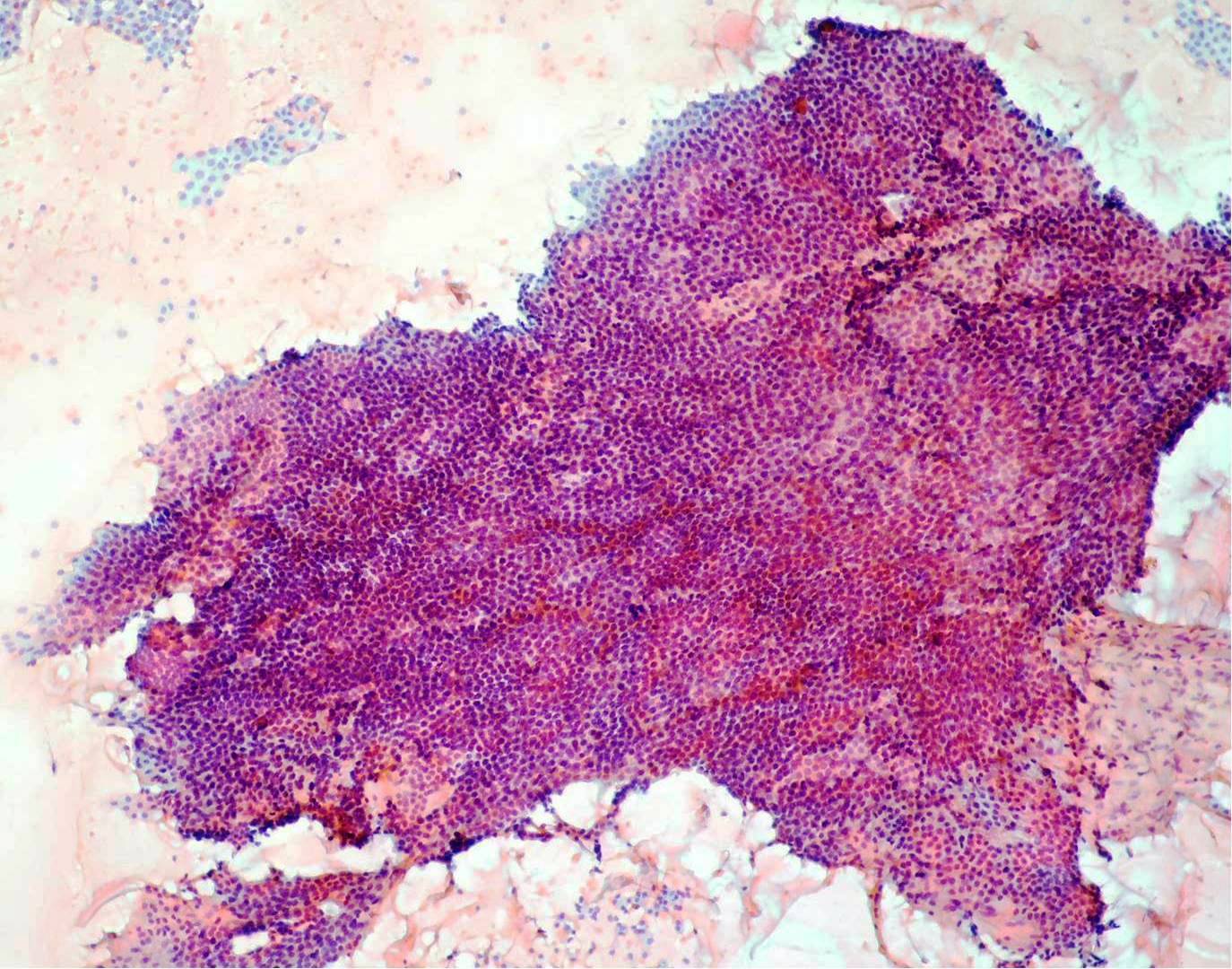
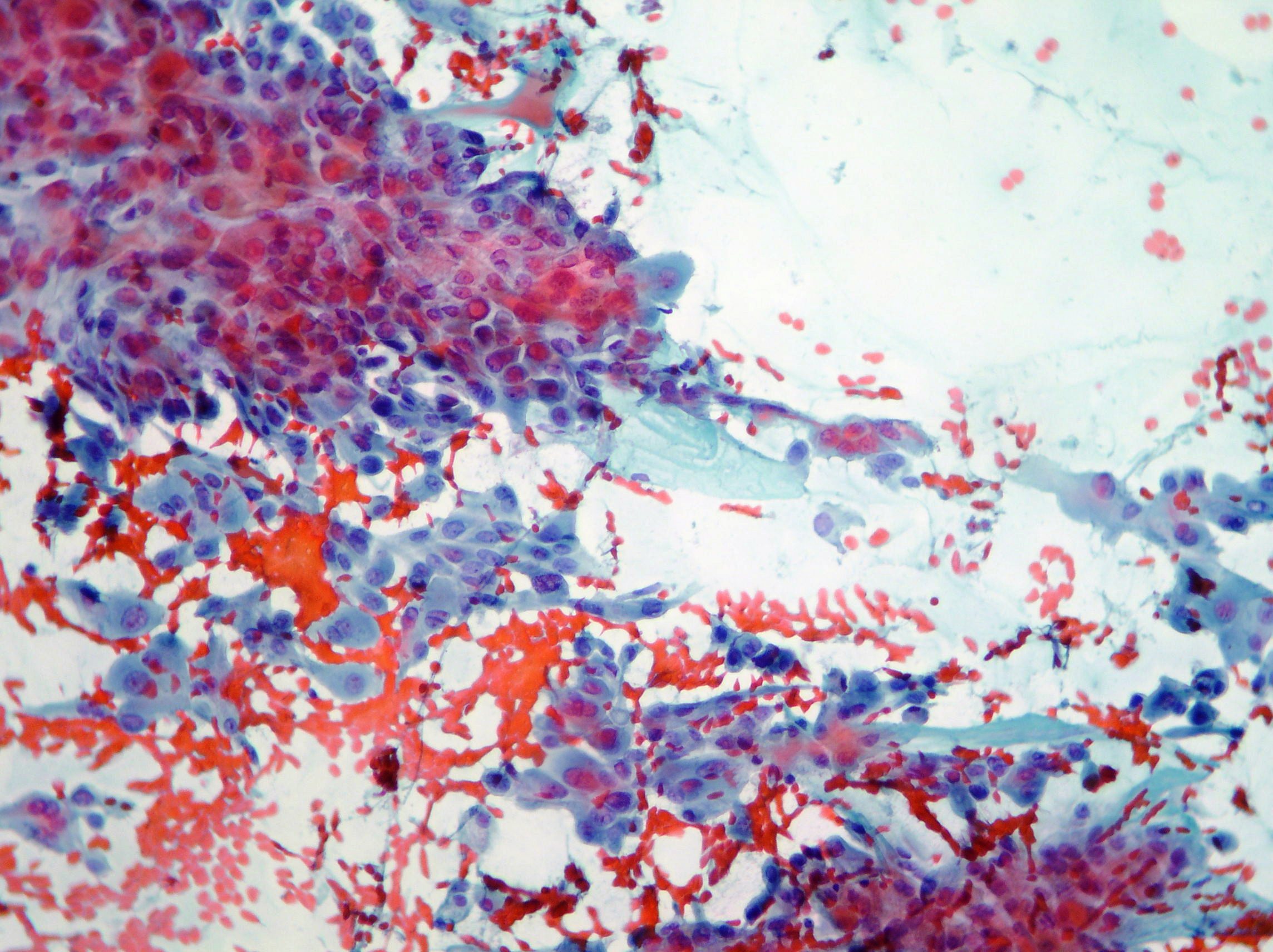
FNAC. Papillary carcinoma
Oncocytic variant of papillary carcinoma showing cytoplasmatic granular eosinophilia associated to papillary nuclear features. (Papanicolaou, x200, x100)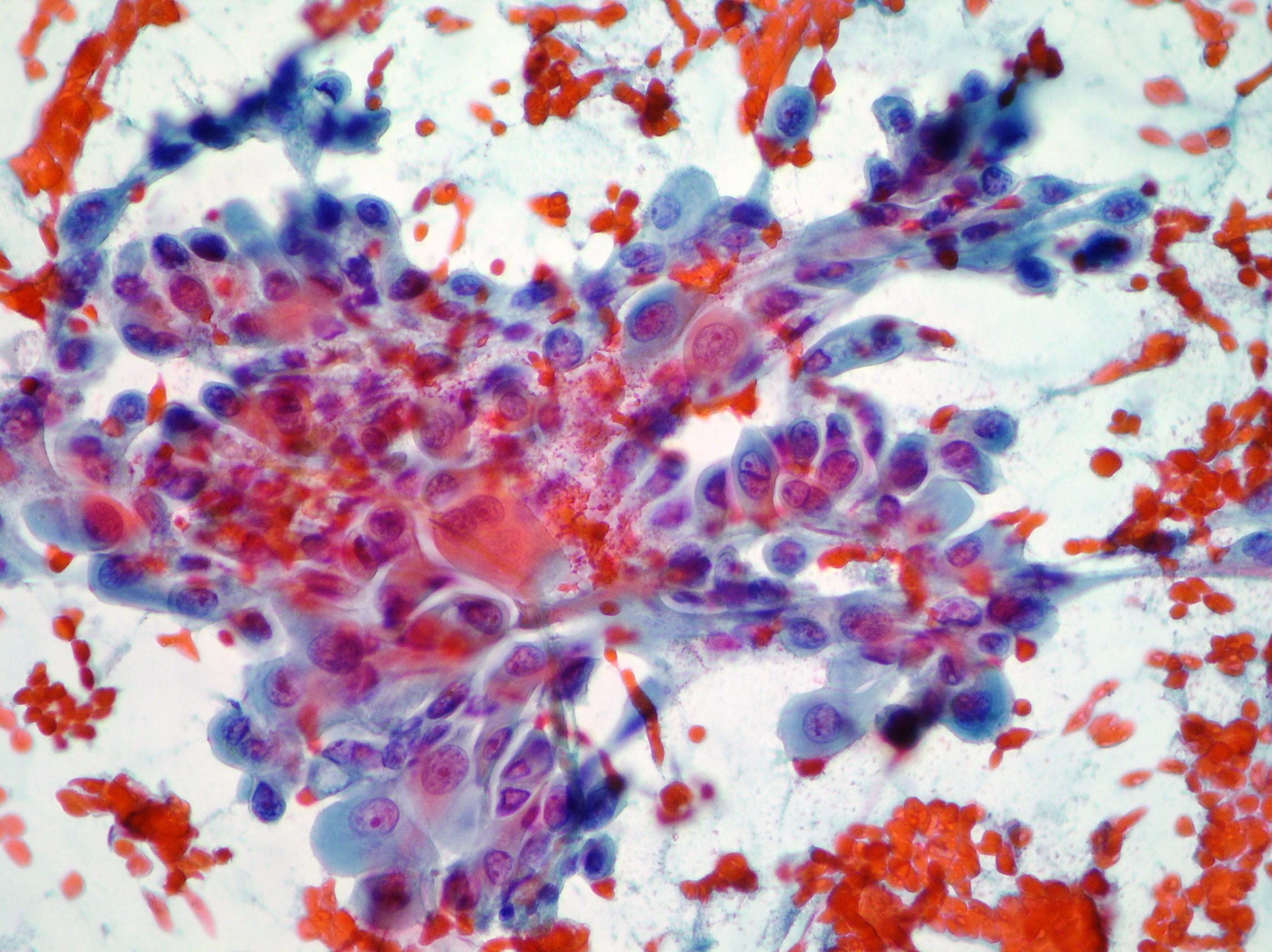
FNAC. Follicular Neoplasia
Cellular smears of thyrocites in microfollicules or rosettes with wreathlike disposition, scant or moderate nuclear atypia, scanty or no colloid in the background suggestive of follicular neoplasia. (Papanicolaou, x200, x100)