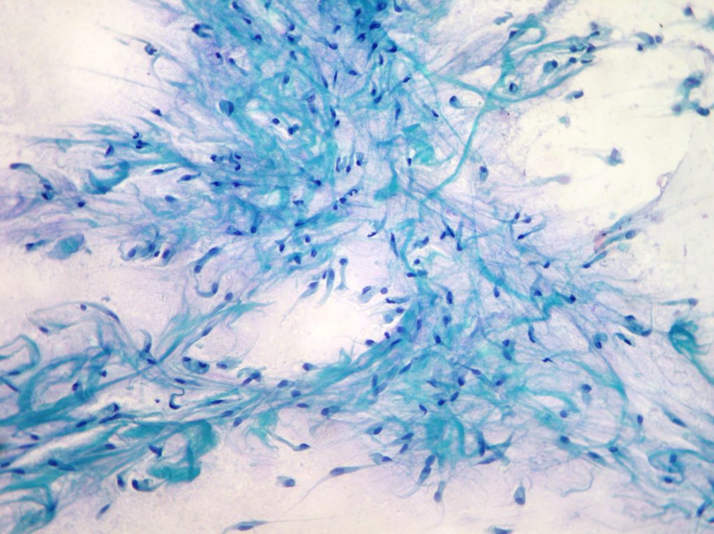
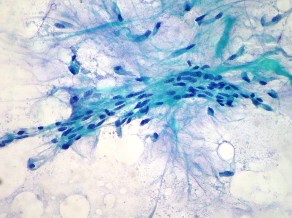
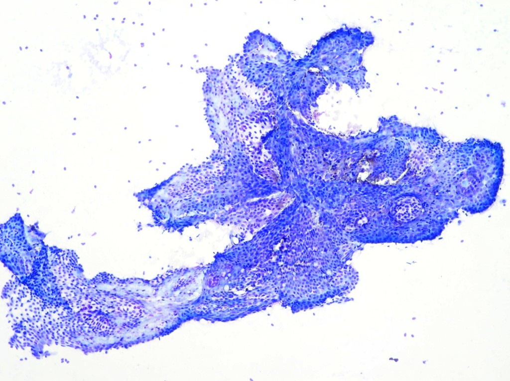
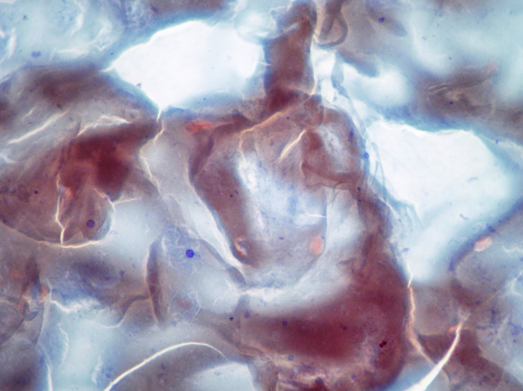
Desmoid fibromatosis of the breast
Breast FNAC showing sheets of bland spindle cells with long fusiform nuclei within matrix. (Papanicolaou x200)
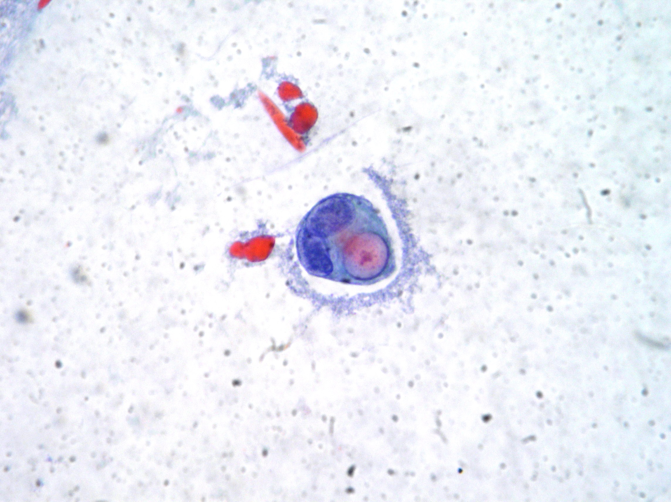
Bone metastasis of lobular breast cancer
Bone metastasis of lobular breast cancer cells showing intracitoplasmic vacuolization with typical targetoid appearance. (Papanicolaou x400)
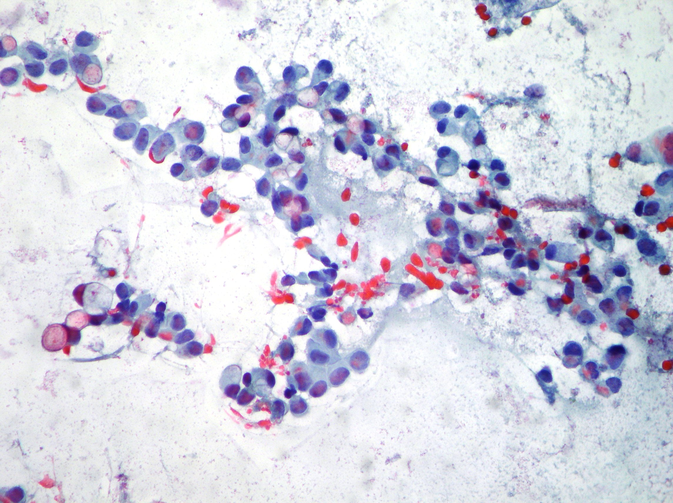
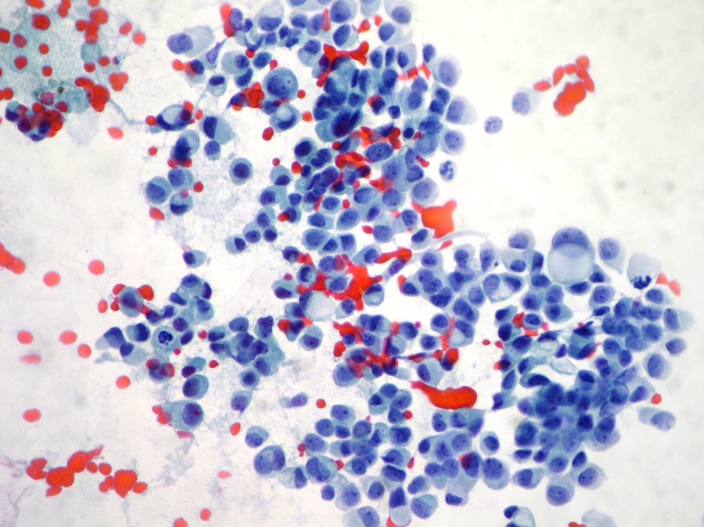
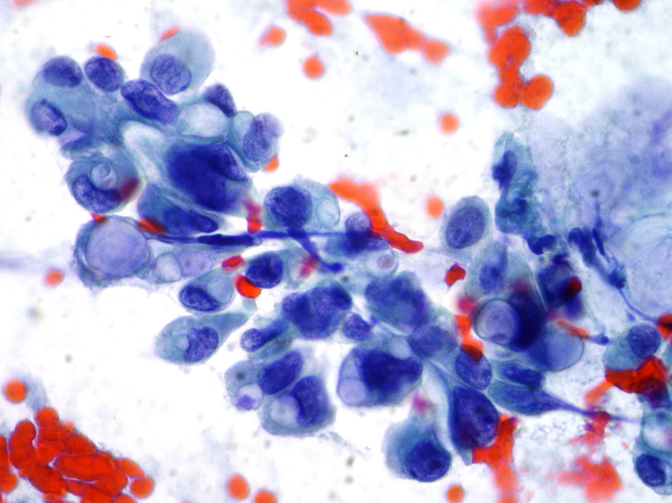
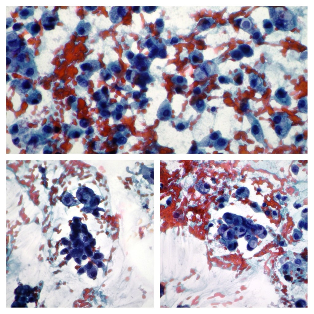
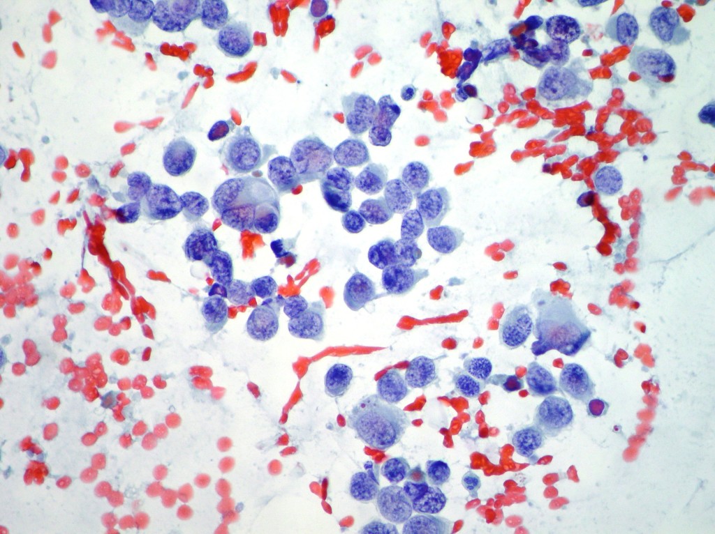
Lobular breast cancer
A case of lobular breast cancer cells showing intracitoplasmic vacuolization with typical targetoid appearance. (Papanicolaou x40, x100 oil immersion)
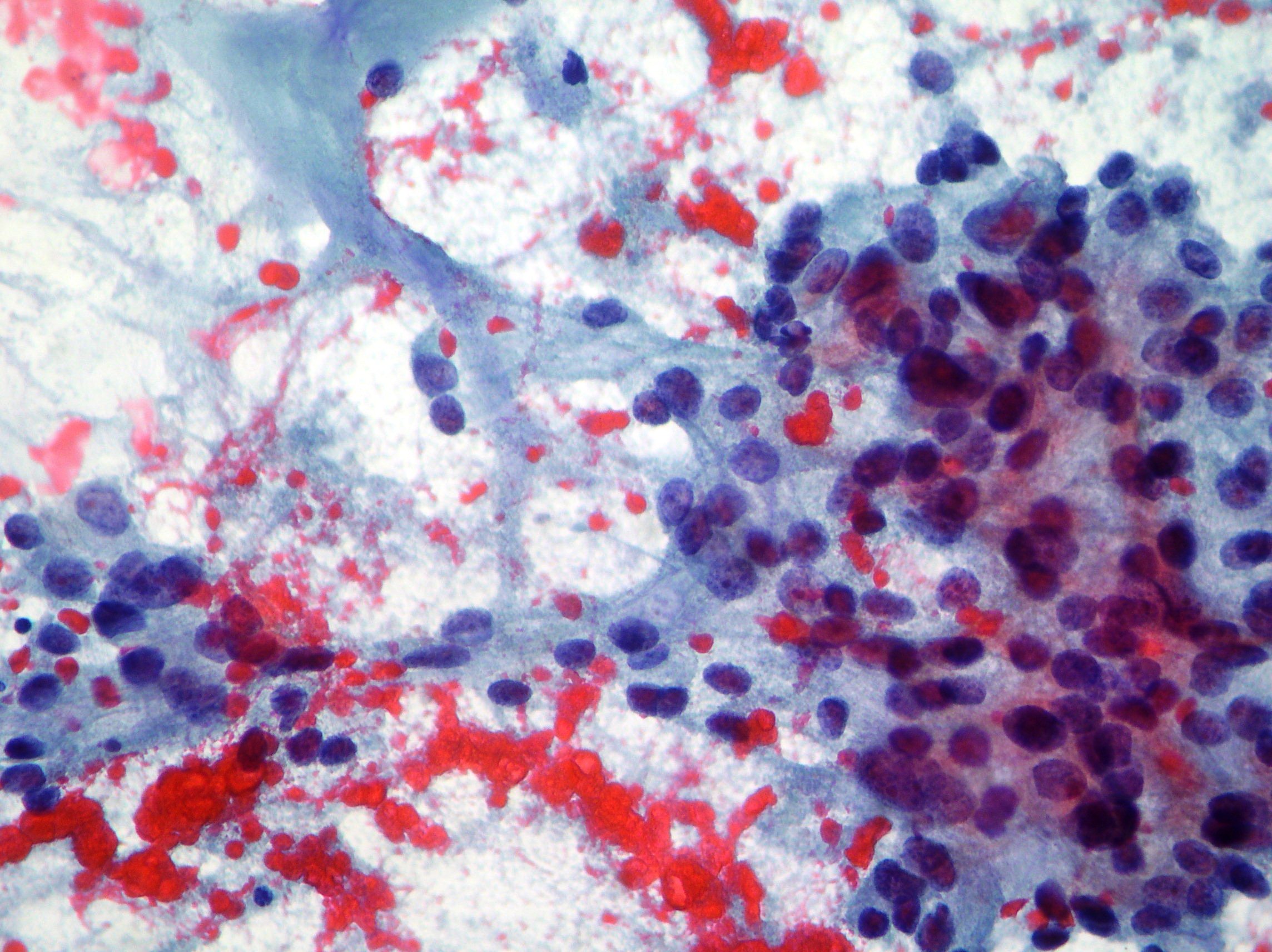
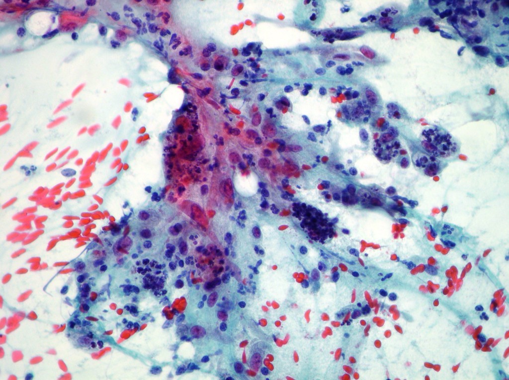
Mucinous breast cancer
A case of mucinous breast cancer showing mucus in the background, elevated cellularity, single ductal cells, three dimensional clusters and nuclear atypia. (Papanicolaou x200)
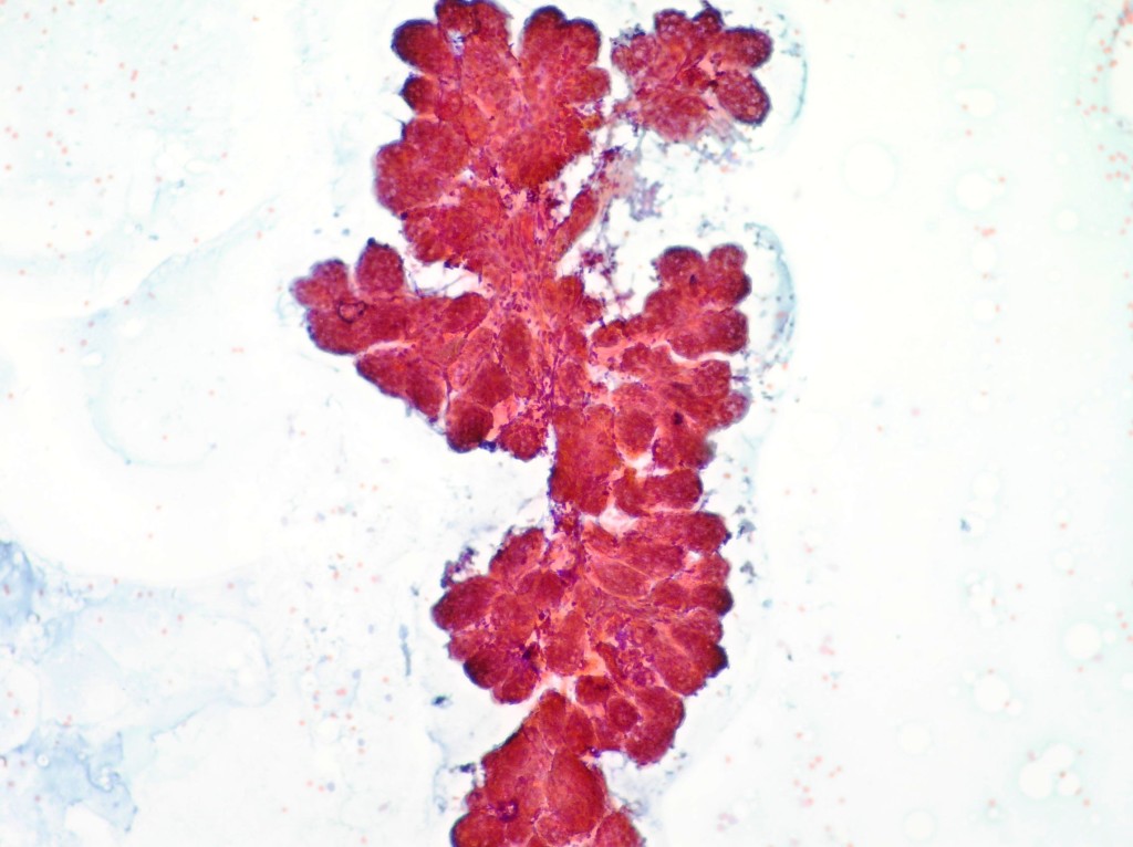
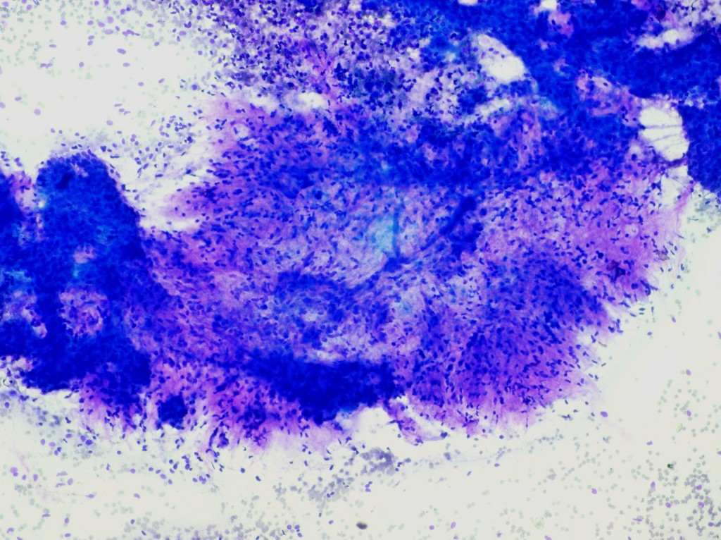
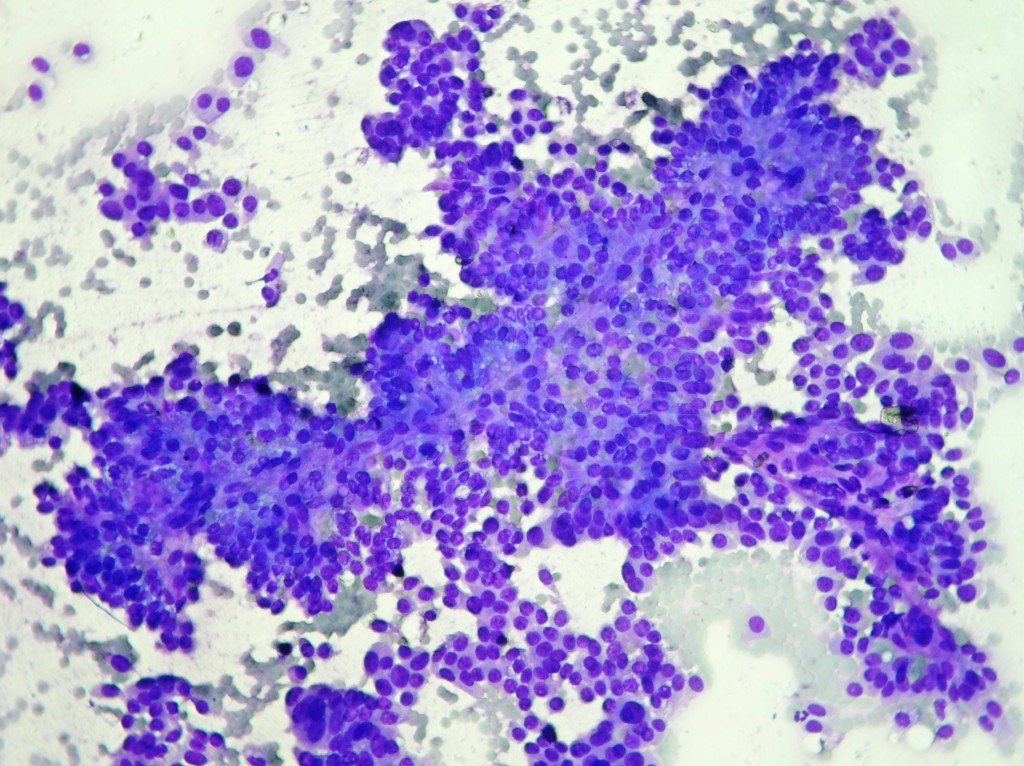
Phylloid breast tumor
Phylloid breast tumor showing predominance of mixoid and cellular stroma over epithelial cells. (Diff Quick ROSE x100)
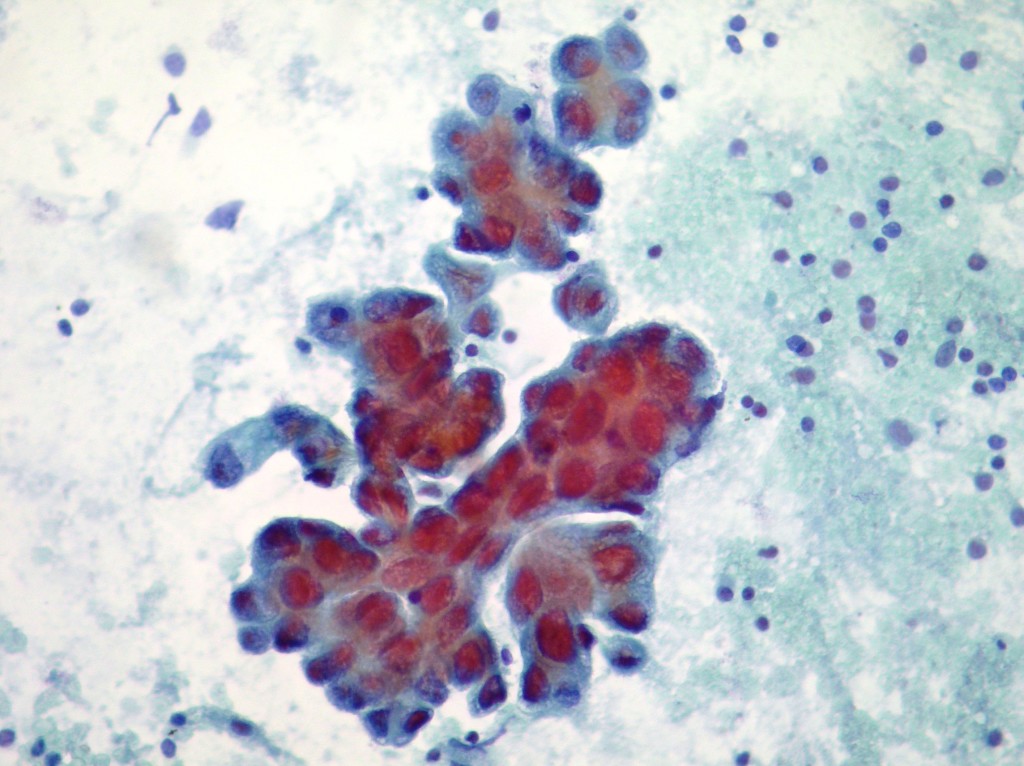
Ductal breast carcinoma
Ductal breast carcinoma cells showing pseudopapillary structures. (Diff Quick ROSE x100; Papanicolaou x200)
Breast ductal cells
Cohesive sheets of breast ductal cells showing benign proliferative features including bipolar nuclei dispersed and over the sheets (Papanicolaou x10, x10, Diff Quick)
Nipple discharge
Serous dense nipple discharge. Rare histiocytes. (Papanicolaou x200)
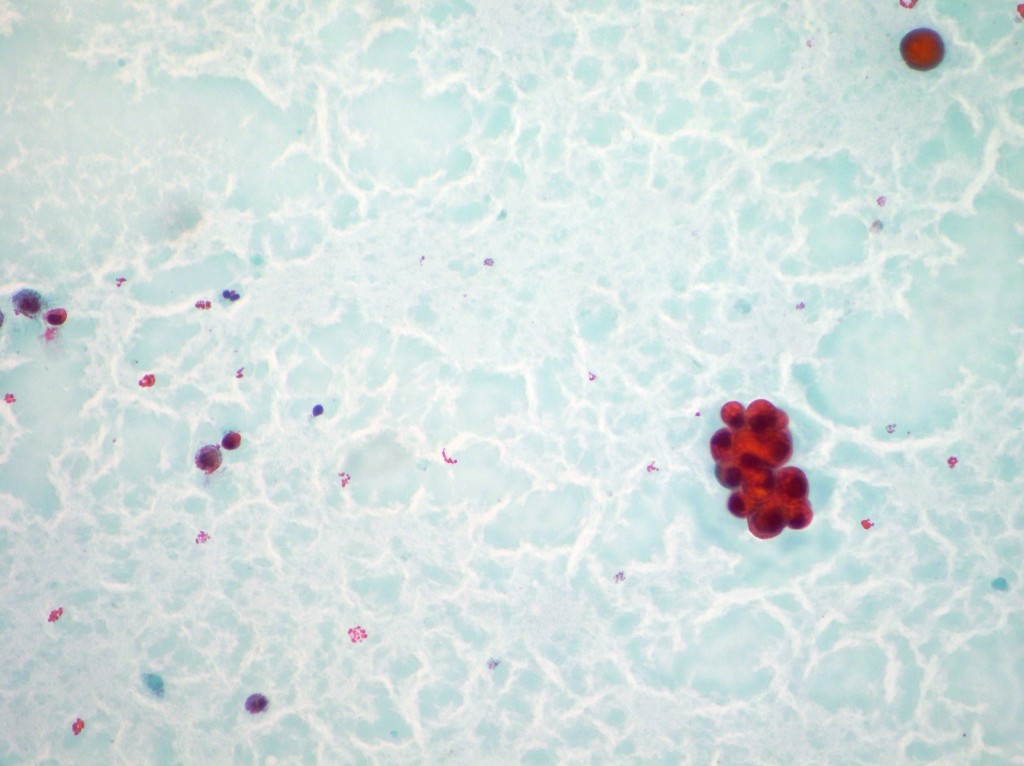
Breast tumor cells
Clusters of breast tumor cells surrounded by extracellular “colloid” material suggestive of mucinous lesion (Papanicolaou x10, x20, R.O.S.E Diff Quick)
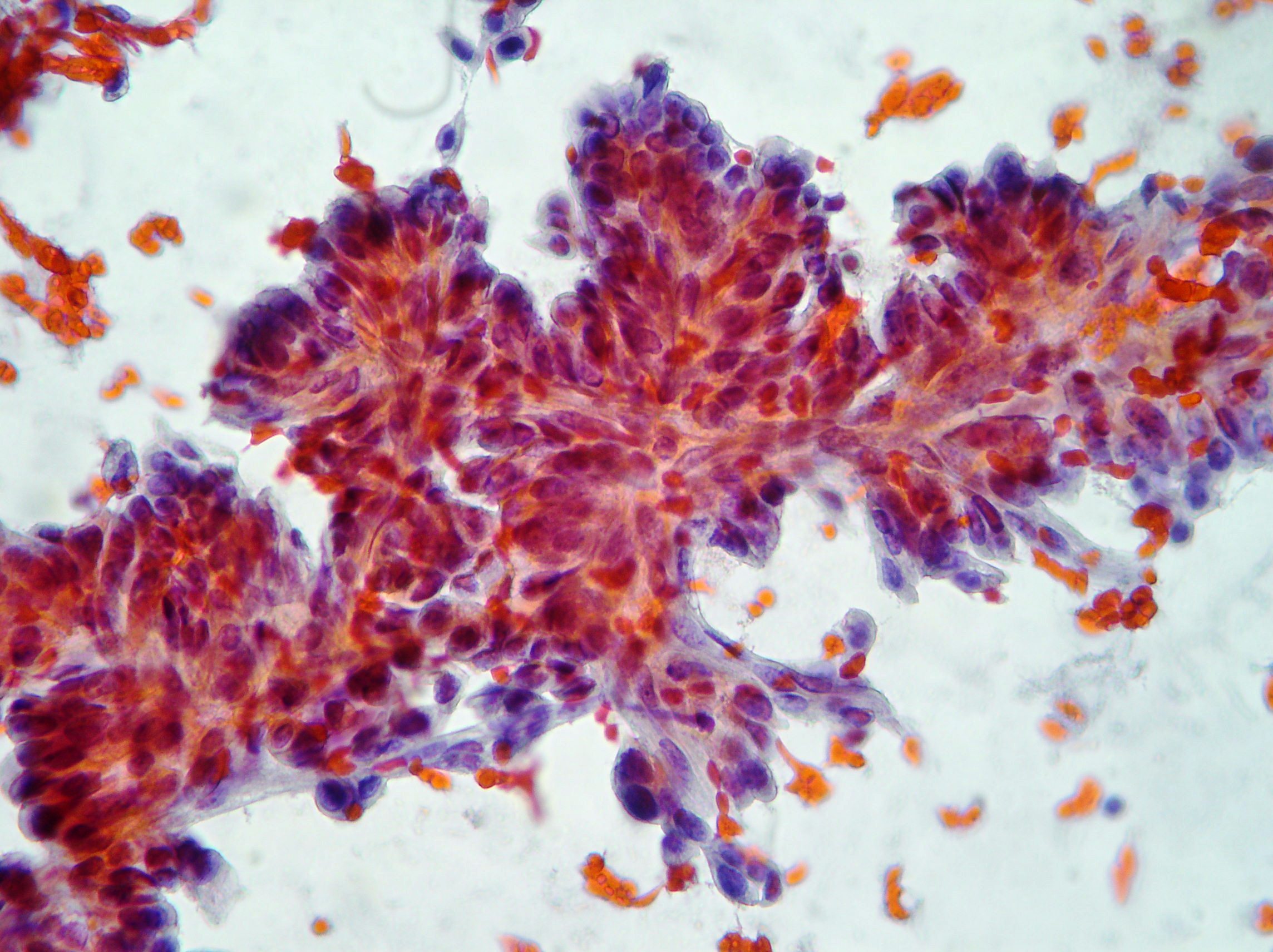
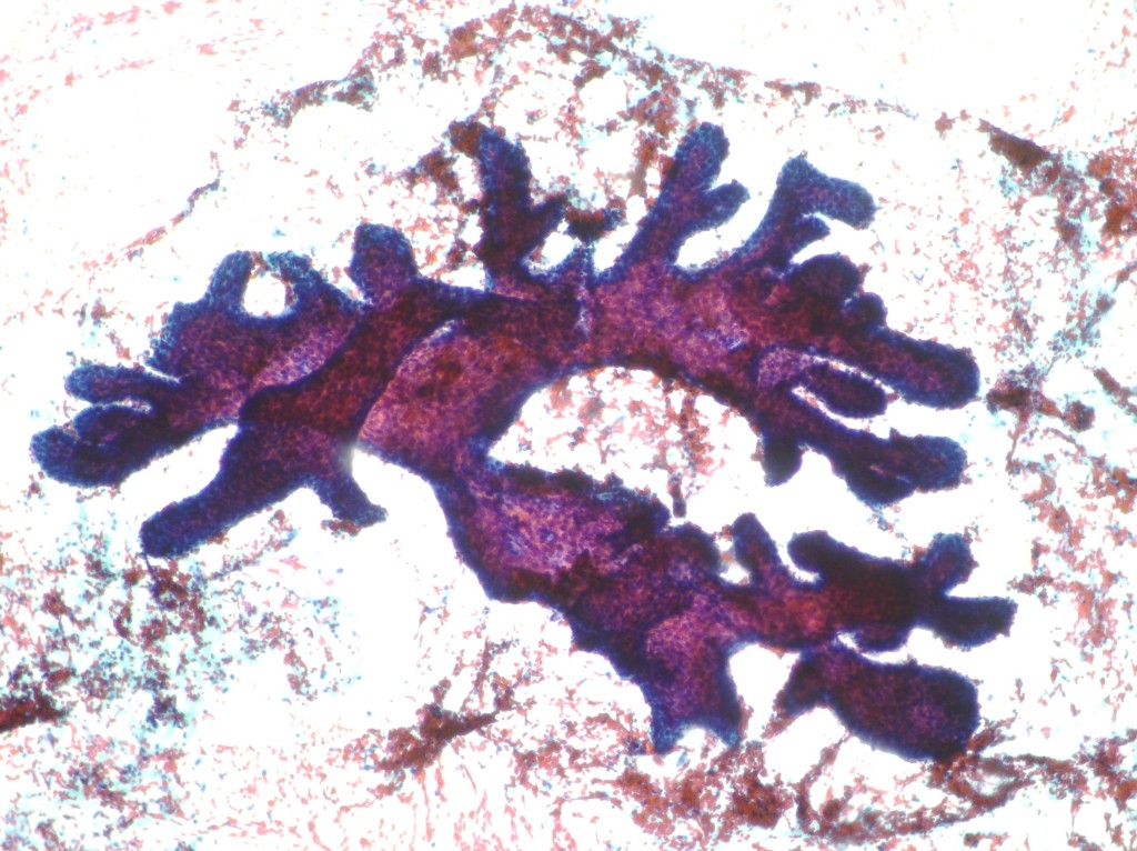
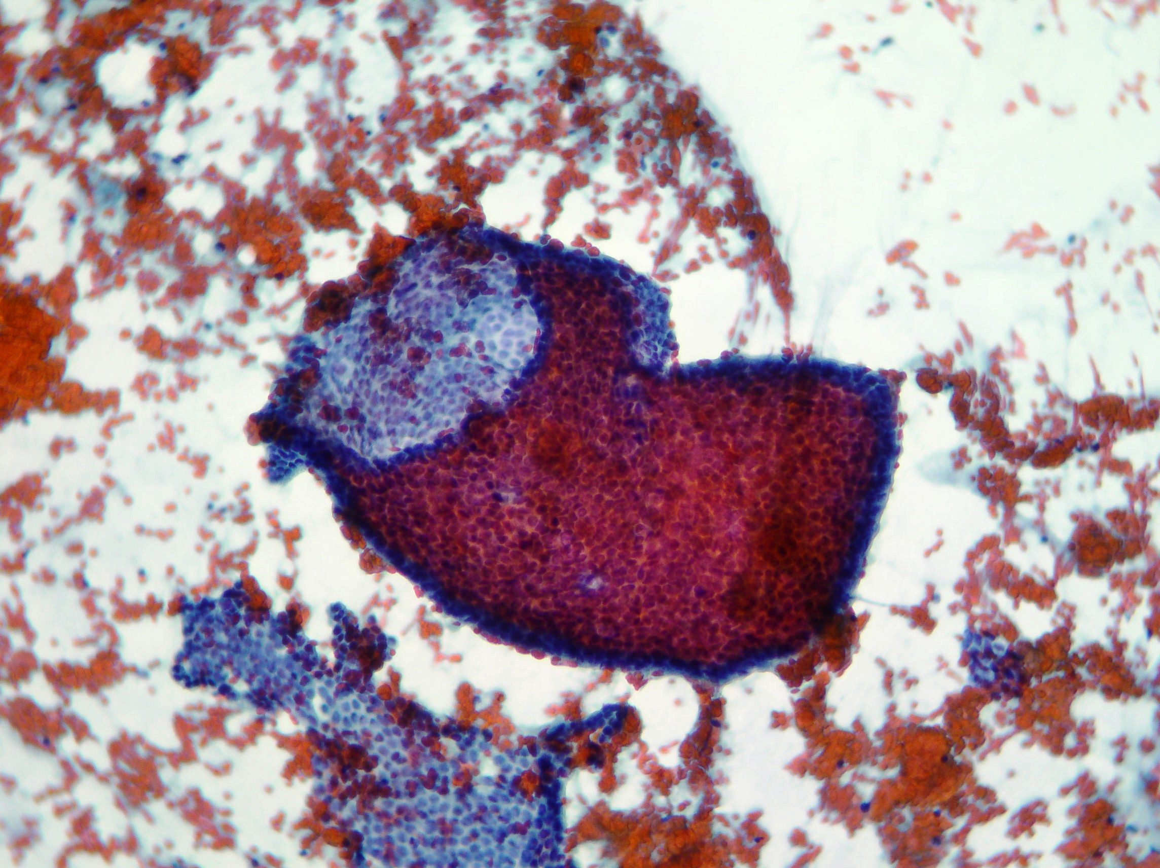
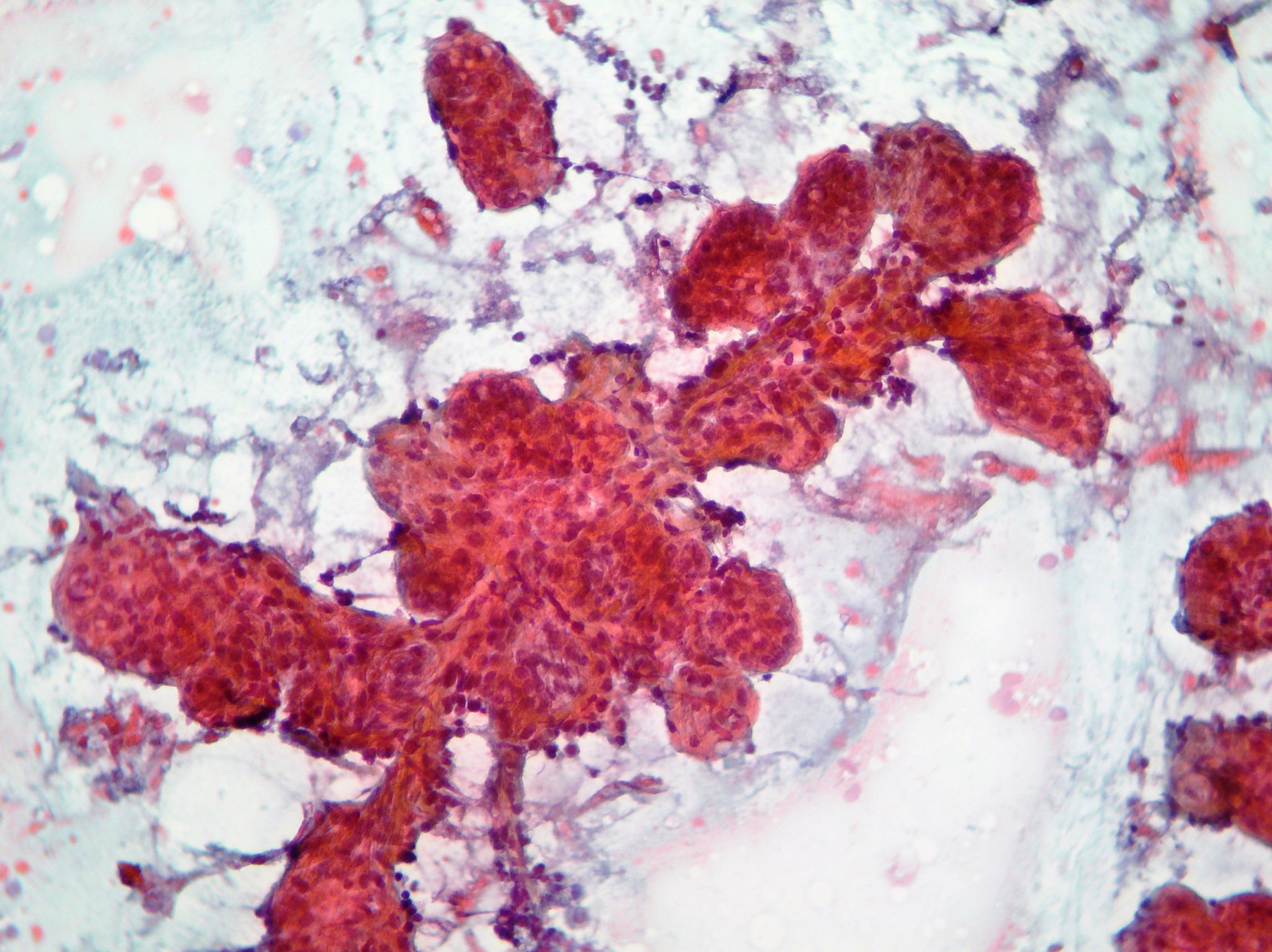
Breast papillary carcinoma
FNAC. Fragment of micro-architecture showing papillae with a fibrovascular core compatible with breast papillary carcinoma (Papanicolaou x10).

Fibroadenoma
Fragment of cohesive epithelial and stromal cells with variable nuclear crowding and overlapping. Single bare bipolar are scattered in the background suggestive of fibroadenoma. (Papanicolaou, x100)
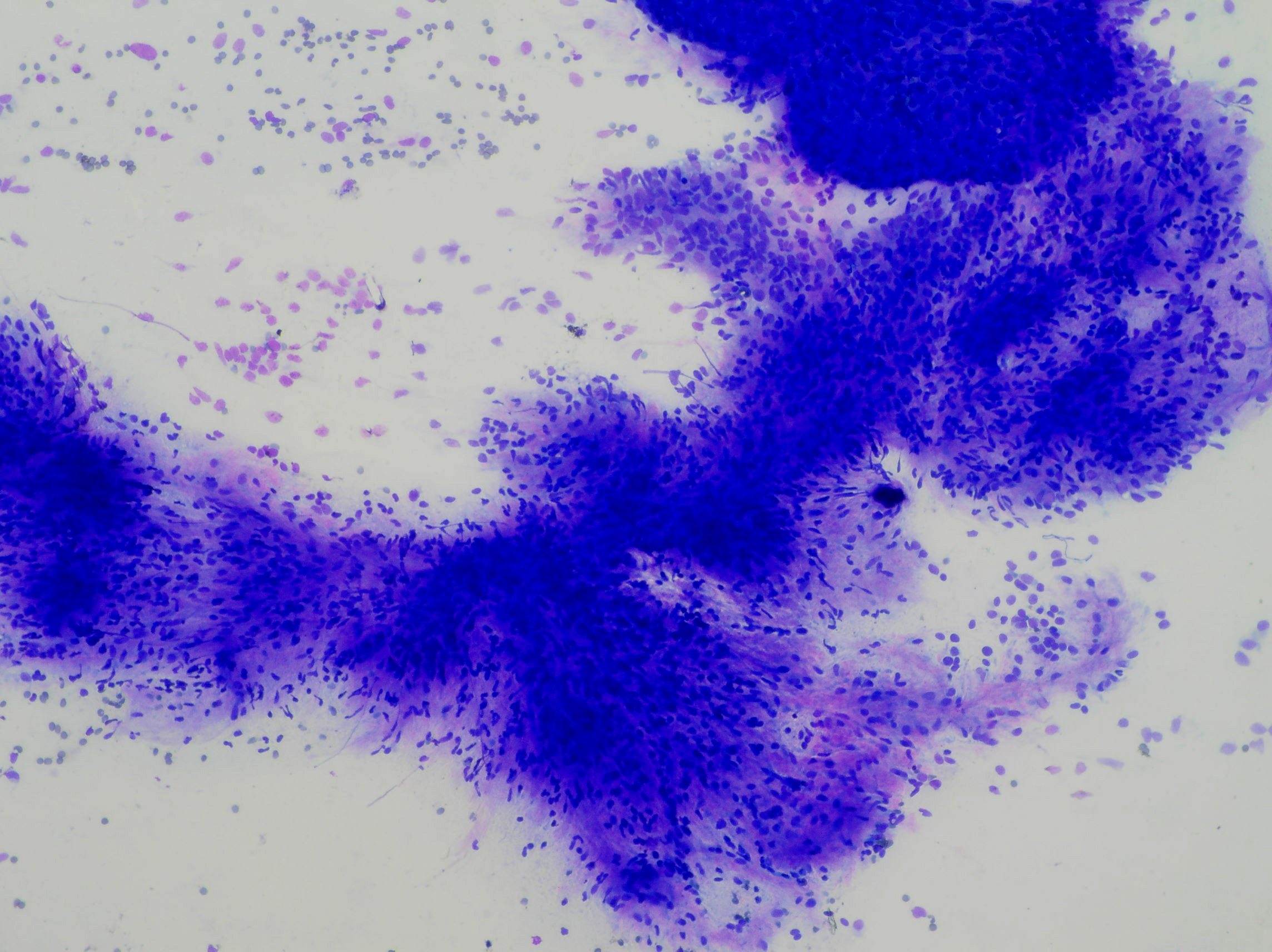
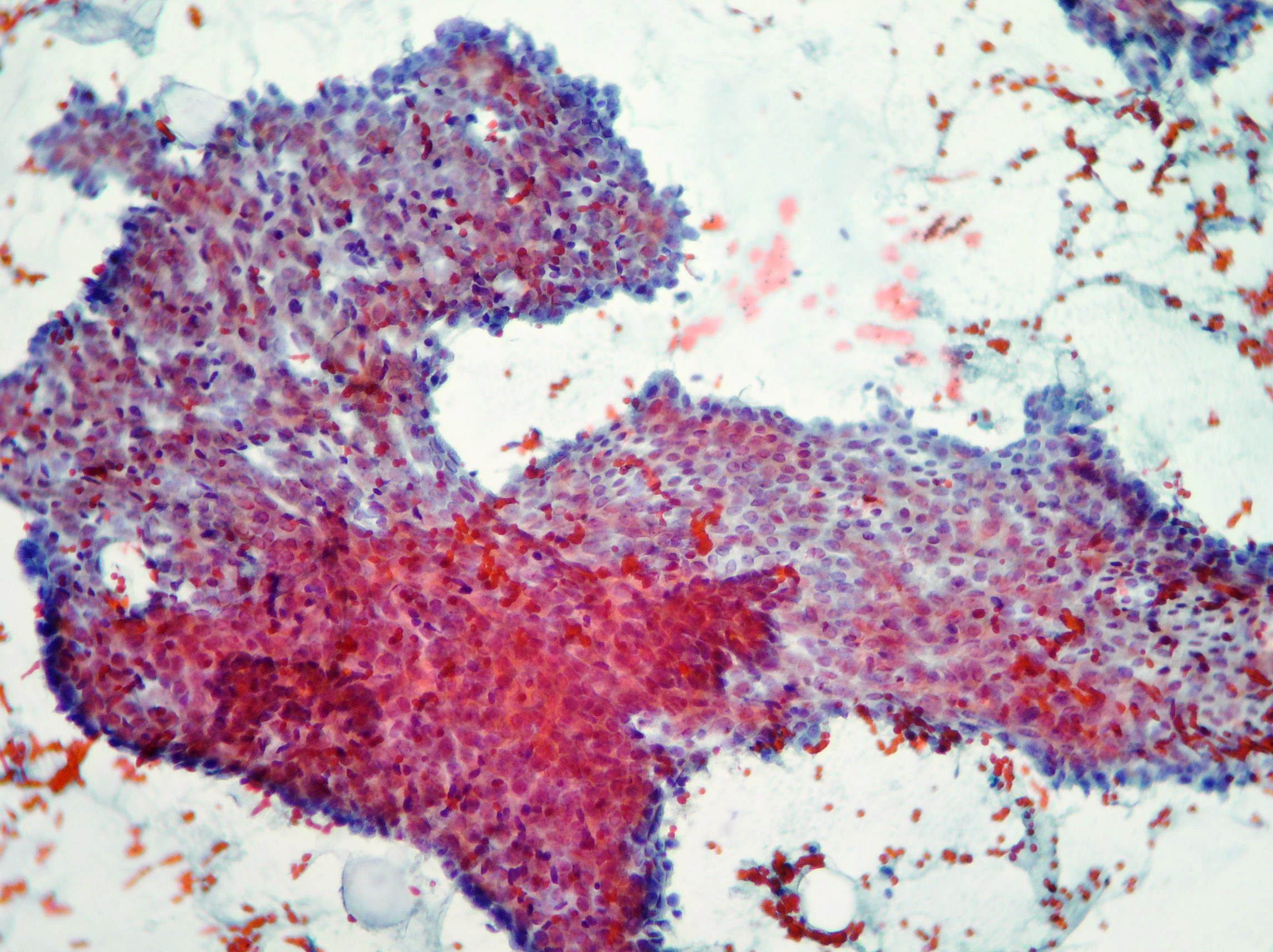
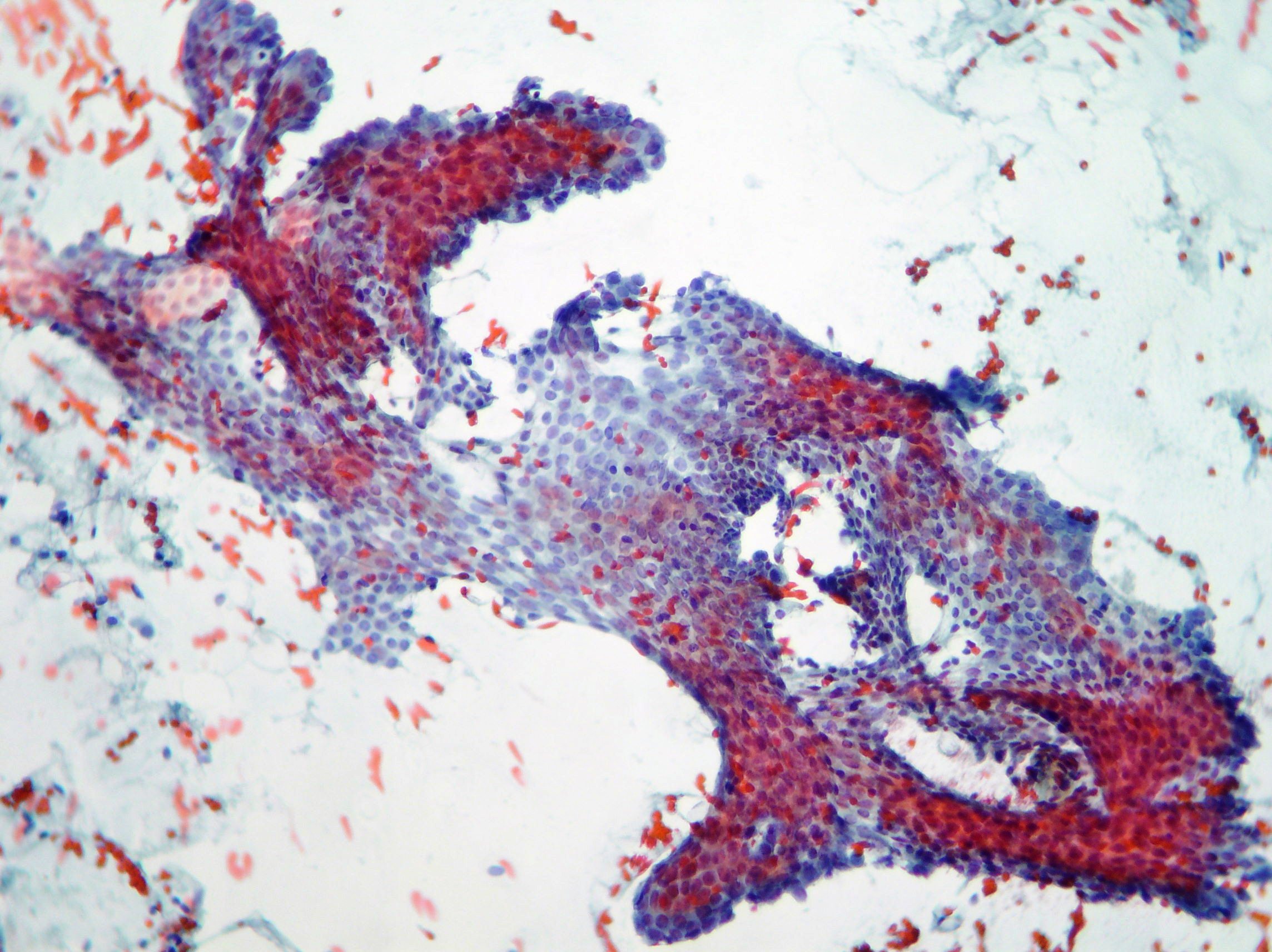
Breast undifferentiated neoplasia
Poorly cohesive malignant cells showing nuclear enlargement and pleomorphism, coarse chromatin and occasional mitotic figure suggestive of breast undifferentiated neoplasia (Papanicolaou x100, x200; Rapid Onsite Evaluation )
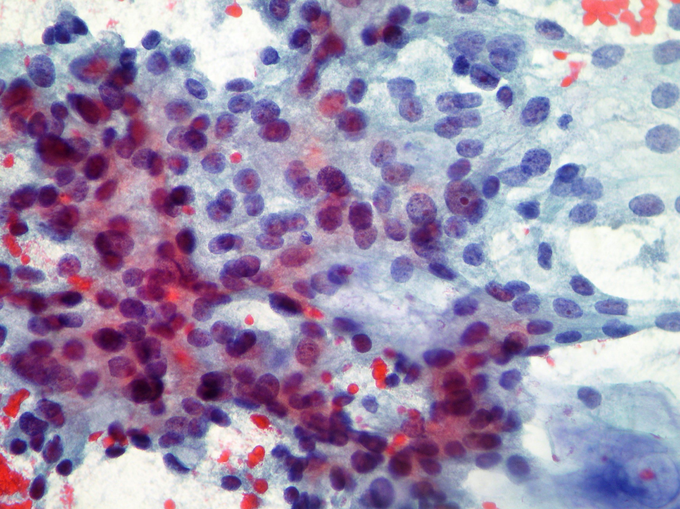
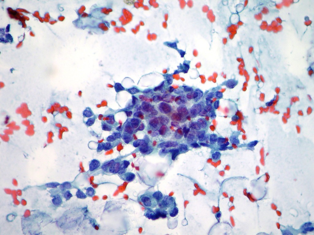
Invasive ductal breast carcinoma
Intermediate grade of invasive ductal breast carcinoma showing poorly cohesive malignant cells, single and in clusters, nuclear enlargement and pleomorphism. (Papanicolaou, x400)
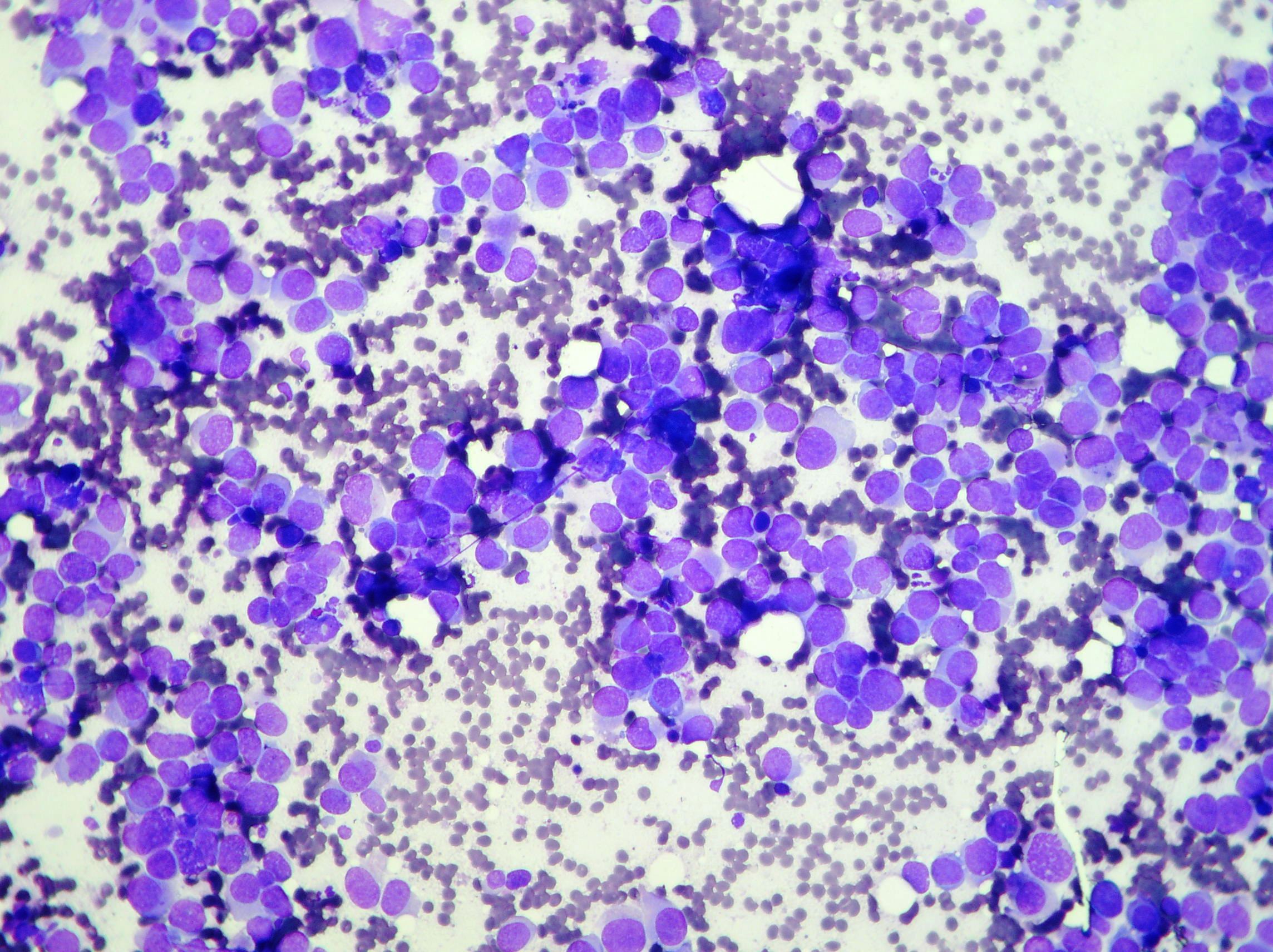
Lymphoproliferative breast disease
Cellular smear composed of dispersed single cells with spherical nuclei and coarsely granular chromatin. Most nuclei contain conspicuous multiple nucleoli. Mitotic figures are present. The case is suggestive of breast lymphoproliferative disease. (Papanicolaou, x200)
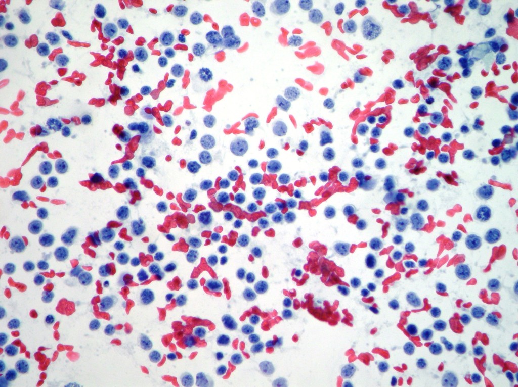
Breast mastitis
Breast mastitis showing neutrophils and epithelioid cells, histiocytes and debris in the background. (Papanicolaou, x200)
Breast apocrine cells
Breast apocrine cells obtained from benign cyst fluid showing a typical aggregate of oxyphilic cells with abundant cytoplasm and enlarged but bland nuclei. (Papanicolaou, x400)
Breast Cystic lesion
Cyst macrophages and a group of epithelial cells in a pseudo papillary structure. (Papanicolaou, x100)
Lobular hyperplasia
Lobular hyperplasia showing numerous acinar cells in lactating patient. (Papanicolaou, x200, x100)